Research Article | | Peer-Reviewed

Evaluation of the Inhibitory Effect and Cytotoxicity of Some Nanoparticles Against Bacterial Isolates from Burn Infections

Received: 4 March 2026     Accepted: 24 July 2026     Published: 17 August 2026
Views:       Downloads:
Abstract

150 swabs were taken from different burns of patients admitted to Medical City Hospital and Al-Kindi Teaching Hospital. 128 swabs showed morphological examinations. The isolates were confirmed at the species level using Vitek2 compact system for skin diseases. The isolates under study were tested for sensitivity using the drill diffusion method. Nanoparticles were prepared by two methods, chemical synthesis and green synthesis, Aloe vera and aqueous extract of saffron (Crocus sativus L.). Bacteria were isolated by 60 isolates of Pseudomonas aeruginosa, (46.87%), followed by Staphylococcus aureus, with 38 isolates (12.28%), Klebsiella spp. with 20 isolates (15.62%), and the rest of the isolates were Escherichia coli, Enterobacter cloacae, Acinetobacter baumannii, Stenotrophomonas maltophilia, with three isolates for each type, i.e. (1.56%). S. aureus and Ps. aeruginosa were the most resistant bacteria to antibiotics, and it was found that substance nanoparticle prepared by Artificial roads nanoparticle. The percentage of Staph. aureus bacteria was (28.12%), while Ps. aeruginosa bacteria was (46.87%). The solutions were at concentrations of (100, 75, 50, 25) M%. It is noted that the highest inhibition concentration was (100) M%. As for the is Staph. aureus and Ps. aeruginosa bacteria isolates zone (48, 45) mm, respectively. It was found that the saffron of (45) mm in inhibiting the isolates of Gram-positive bacteria Staph. aureus. The success of Staph. aureus with nickel oxide with saffron at a rate of MIC. Invivo test animals, and the MBC the conducted two methods tests were, ZETA POTENTIAL. Nanomaterials with a smaller size of 5-12nm called quantum dot were observed, and no agglomeration occurred in them. It was shown that the MTT toxicity assay for the solutions of the IC50 ratio for cancerous PC3, while for cellsand the substance is not toxic to the solution for treating S. aureus bacteria, and Ps. aeruginosa.

Published in International Journal of Clinical and Experimental Medical Sciences (Volume 12, Issue 3)
DOI 10.11648/j.ijcems.20261203.11
Page(s) 39-56
Creative Commons

This is an Open Access article, distributed under the terms of the Creative Commons Attribution 4.0 International License (http://creativecommons.org/licenses/by/4.0/), which permits unrestricted use, distribution and reproduction in any medium or format, provided the original work is properly cited.

Copyright

Copyright © The Author(s), 2026. Published by Science Publishing Group

Keywords

Aloe vera, Pseudomonas aeruginosa, Staphylococcus aureus, Crocus sativus L, Nanoparticles

1. Introduction
Burns are skin injuries resulting from exposure to heat such as (fire, hot liquids, or gases, electricity, chemicals such as strong acids and bases, or exposure to radiation such as (ultraviolet rays, X-rays or microwaves), proper management of burns reduces the risk of complications resulting from burns and in order to prevent infection, new therapeutic methods are needed. Nanotechnology has provided a group of molecularly designed nanostructures that can be used in all therapeutic applications for burns . Molecularly designed nanostructures can be divided into organic and inorganic such as polymeric nanoparticles and silver nanoparticles, in addition to healing approaches based on nanotechnology. Technological development in the field of Nano medicine is useful in finding new alternatives. Several types of nanoparticles have been proposed, which have been used to eliminate antibiotic-resistant bacteria. Skin infections are diseases that medicine relationship human appearance. Its external appearance and health, and these infections occur after the pathogen is able to overcome and penetrate the skin, which is the first mechanical barrier, causing skin infections, which vary in terms of their depth and the type of microscopic organism causing them. There are bacterial, viral, fungal infections, and others . Many medicinal plants are used to treat skin, viral, and fungal infections. Many antibiotics are also used to treat the causes of these infections. However, due to the frequent random use of these antibiotics, many bacterial and fungal isolates have spread that are resistant to the action of these antibiotics . The Staph. aureus species is characterized by its ability to ferment mannitol sugar and its resistance to high salt concentrations, which reach (7.5% table salt), in addition to giving a complete blood analysis. haemolysis β. It is present It is naturally found in the human body as it is considered one of the natural flora in the upper respiratory system, especially the nasal area, and it is also found on the skin, specifically in moist places and folds, and enters the tissues through scratches, wounds and burns . By penetrating the surface of the skin, it causes boils loaded with abscesses in the hair follicle, forming folliculitis. When it enters deeper tissues, it causes carbuncles that exude pus to the surface of the tissues and are hardened and painful with a reddish circular shape and often spread on the neck and require a long period to heal . In a study conducted by on bacterial skin infections, it was found that Staphylococcus aureus constitutes 60% of all bacterial skin infections. These bacteria possess virulence factors, which are molecules present or secreted by the bacteria that play important and multiple roles in pathogenic infection, such as colonization of host cells, adhesion to cells, invasion and inhibition response. There are a number of virulence factors possessed by Staph bacteria. aureus, which plays a role in its pathogenicity . Virulence factors include the adhesion factor that provides food for bacteria, cell surface proteins that form the adhesion template for bacteria , the presence of protein A, which is an anti-phagocytic surface protein , the presence of cilia, which are structures found on the surface and are important because they mediate the adhesion process, the presence of the capsule that stimulates virulence by obstructing the phagocytosis process, the presence of teichic acids, which are negative ionic polymers, and their functions include maintaining the structure, integrity, and shape of the bacterial cell and contribute to many vital activities such as resistance to environmental conditions and lytic enzymes, and contribute to cell division and morphogenesis . In addition, there are extracellular enzymes Extracellular enzymes that resist stress such as catalase, protease, nucleic acid-degrading enzymes, hyaluronidase, which contributes to the spread of toxins in tissues, lipase, which is important in nutrition and spread, staphylokinase, which is important during bacterial cell division in the late phase of division, and there is also coagulase, which is a plasma coagulase enzyme and is important in diagnosis . Some types of staphylococci secrete a group of exotoxins such as nuclease, protease, lipase, coagulase, and others . The exotoxins are Alpha-hemolysin, Beta-hemolysin, Gama-hemolysin, Leukocidin, Panton-valentin, Toxic shock syndrome Toxin, and intestinal toxins . Pseudomonas aeruginosa is rod-shaped not form spores, motile and is aerobic. Ps. aeruginosa causes are difficult to treat due to its multiple resistance to antibiotics . powerful such diverse its hemolysin and proteinase, to its production of many exotoxins that are as effective as diphtheria toxin . It also has the production of broad-spectrum beta-lactamase enzymes . They are well-established in the moist environment of hospitals, as they have been isolated from beds, soap containers, and disinfectant solutions, and through them they move to wounds and burns, causing necrosis of these areas, tissue destruction, and the exudation of blood plasma outside the skin to become a good breeding ground for bacterial growth. These bacteria cause pururitic maculopustulur and vesiculopustulur rash, which is associated with swimming in contaminated pools. They also form biofilms, which are biofilms consisting of colonies of microorganisms surrounded by exopolysaccharides produced by the bacteria ps. aeruginosa, in addition to the pigment pyocin and functional proteins, all of which contribute to building the biofilm . The presence of biofilms to protect these bacteria from harmful environmental factors and the presence of membrane tissue material is one of the main resistance mechanisms in Pseudomonas aeruginosa biofilms. . The resistance to multiple antimicrobials shown by a type of microorganisms towards a spectrum of antibiotics and multiple treatments, including fungi to many antimicrobial drugs, and these strains can be comprehensively resistant . Distinguishing the types of resistance Resistant bacteria that pose a risk of transmitting infection and rapid infections, as they infect patients in hospitals and intensive care units, or are cyclic resistance, and the infection caused by these resistant strains is accompanied a long period of treatment . From what was presented, the aim of the study was to try to know the effect of biosynthetic nanocomposites on the characteristics of pathogenic bacteria isolated from burn infections, especially those with multiple resistance for antibiotics.
2. Materials and Method
NiO and Cobalt oxide nano solutions were prepared. 4% sodium hydroxide was prepared and added to nickel nitrate solution (Ni(NO3)2) 29% M. The temperature was gradually raised to 70°C (Al-Sawy, 2017). (Saraev, 2005) NiO GREEN NANPRACTICAL was prepared. 1% M was prepared in the same way. Nickel nitrate solution (Ni(NO3)2) was prepared (Joseph, 2012). Nano cobalt oxide CoO was also prepared by chemical method to prepare nano cobalt oxide NANOPARTICAL STRACTUR by preparing sodium hydroxide NaOH 4% to which cobalt nitrate solution (Co(NO3)2) 37% M is added. The two solutions are mixed in the same previous way. Cobalt oxide is also prepared with nano Aloe vera solution, where 10% of Aloe vera extract is prepared and add to the cobalt nitrate solution (Co(NO3)2) 37% M and mix in the same way as before Then mix the two solutions, nickel oxide and cobalt oxide solution, and mix them percentages Place in a vibrating device to mix two materials until 50 (± 3) in a water bath. The culture media are prepared and the collected swabs are cultured and the bacteria are diagnosed using Vitek2 compact chemically and morphologically .
3. Results and Discussion
3.1. Bacterial Examination
The strains that have become resistant to all available therapeutic agents, including resistant to inhibit the production of penicillinase by S. aureus, but it has resistance to a number of other antibiotics, aminoglycosides, macrolides, tetracycline and chloramphenicol Vancomycin. The reason for the resistance is the excessive use of an mechanism by bacteria through many defense mechanisms, as well as the lack of healthy practices to combat infection, and the lack of responsible supervision of antibiotics so that they are widely used to avoid increasing resistance . Bacteria can produce enzymes that have the ability to break down antibiotics called beta-lactamase such as β-lactamase. The beta-lactam enzyme is divided into four enzyme classes by producing chloramphenicol acetyl transferase Chloramphenicol acetyl transferase . Some organisms show a mechanism of resistance to the beta-lactam enzyme by an antagonistic effect, which is the synthesis of as they resist tetracycline, which Efflux pumps in the bacterial cell, where their function is to remove waste and can remove antibacterial drugs by means of the pumps .
Table 1. Effect of different concentrations of nanomaterial on inhibiting bacterial growth of cobalt oxide.

The method

Concentrations

Average length diameter of inhibition zone mm

M%

Ps. aeruginosa

Staph. aureus

N1

25

0.0

0

50

0.0

25

75

0.0

13

100

14

11

N2

Aleovera

25

0.0

0

50

35

35

75

37

35

100

47

45

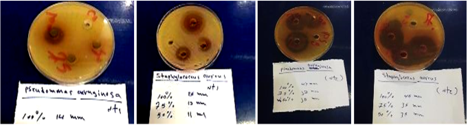
Figure 1. Aloe vera plant extracts with cobalt nitrate on bacterial growth. Zones of inhibition on culture media using NT1 and NT2.
3.2. Nanomaterials XRD Test Results
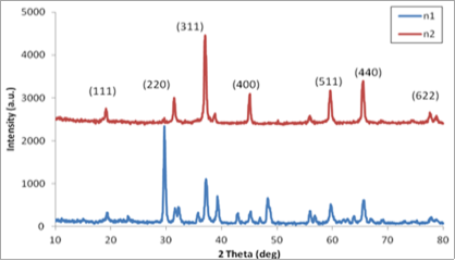
Figure 2. The XRD graph NT1, NT2.
The presence of nanomaterial 19 (111), 31 (220)), 45 (400), 59 (511), 65 (440), 79 (622) in each of the materials NT1, NT2, and the results match the graph of NT1, NT2 with the international measurements of the Miller coefficient and theta degree 2 Theta (deg)).
3.3. SEM Examination
Figure 3. The graph showing the SEM, NT1, NT2.
The results, as shown in Figure 3, represent a substance deposited on glass, where the background appears black. The particle shapes are flower-shaped with dimensions not exceeding 200 nm on the drawing scale, and these particles appear with dimensions on the drawing scale of 50-90 nm, smaller than 100 nm.
3.4. TEM Examination
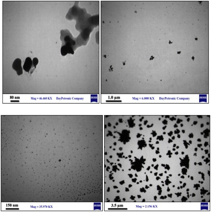
Figure 4. TEM Cobalt oxide solution NT1 Cobalt oxide solution NT2.
It was noted from Figure 4 that, Figure 4, NT1A was 3.5 µm, and according to the scale in Figure 4, NT1B, the nanoparticle 60 nm. . that the of Figure 4, NT2A was 3.5 µm, are single and large in Figure 4, NT2 B, the nanoparticles appeared, the drawing scale was 150 nm, clusters are observed, and the nanoparticle does not exceed 0.5 nm.
3.5. UV Examination
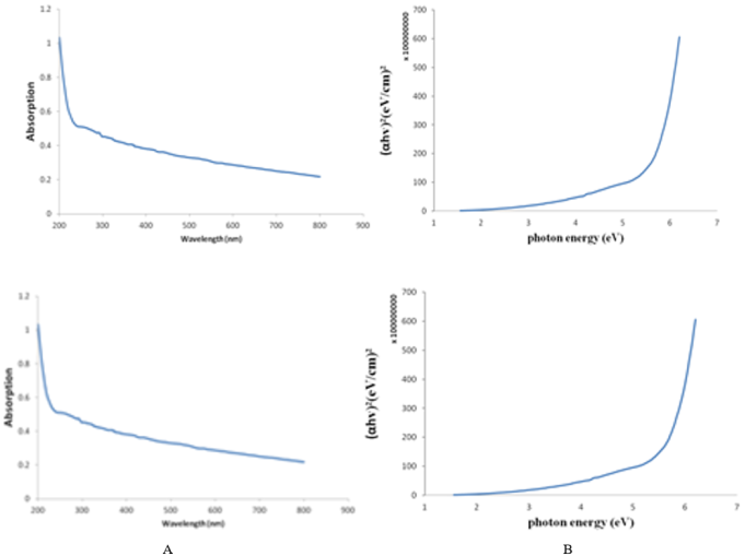
UV examination is wavelengths from 200-400 nm are called ultraviolet rays and are symbolized by UV, while rays from the range 400-700 nm are called visible rays (VIS), and strong rays at a wavelength of 700 nm are near-infrared rays (N-Infra).
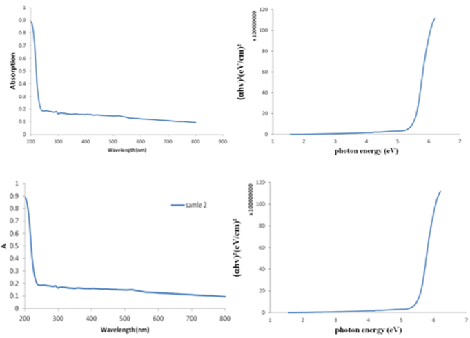
Figure 5. UV, gap, absorbance NT1, NT2.
It is noted from Figure 5A UV that graph shows value is 3.5 ev and nano material. As for Figure 5B UV of NT2 graph A transmittance low absorbance in 236-800 nm. This is attributed the B graph has a 3.8 ev, and it achieves the nanomaterial.
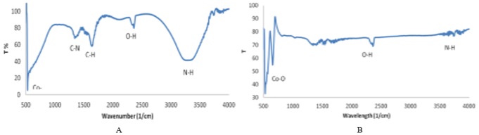
Figure 6. FT-IR, NT1, NT2.
3.6. FT-IR Examination
FT-IR examination It is noted from the graph, Figure 6, A FT-IR of the NT1 cobalt oxide solution that the sites of the bonds Co-O, its presence is observed, while 1315 level there C-N bonds, 2400 O-H bonds, 3251 N-H bonds. It was also noted from the graphic Figure 6,B, FT-IR of the NT2 cobalt oxide solution, that material C, O, H bonds mineral materials, metal and absence of acidic materials, also oxidation and reduction the change the process clumping does not occur. It is noted that whenever the nanoparticle material is diluted, does not clump and sinks to the bottom of the container during preparation.
3.7. Zeta Potential Test
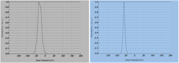
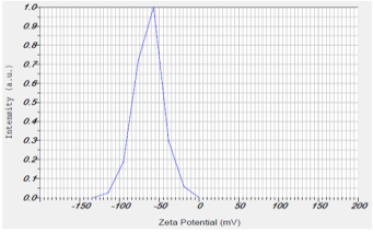
Zeta potential test It was noted from the results of the Zeta test shown in Table 2 that there is a positive repulsive is an indication while are repulsive the prepared nano of cobalt oxide had the value of the Zeta Potential graph, Figure 7, which was -57.3 was negative were low, indicating that no agglomeration occurred in the nano material. As for the nano solutions N2 value of the graph was -28.5 was negative were low, indicating that no agglomeration occurred in the nano material .
Figure 7. The graphs of the results of the zeta potential test for the nanocomposites samples and the samples of the two nanomaterials.
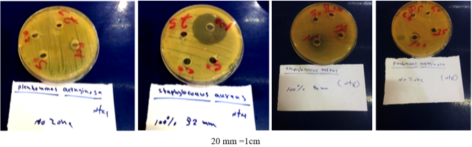
Table 2. ZP results for the nanocomposites samples and the samples of the two nanomaterials combined.

Peak No.

Zeta Potential

Electrophoretic Mobility

N1

-57.3 mv

-0.000444 cm2/vs

N2

-28.5 mv

-0.000221 cm2/vs

Table 3. The effect of different concentrations of nickel oxide nanomaterial on inhibiting bacterial growth.

Type of discriminatory material

Average length diameter of inhibition zone mm

Concentrations M%

Pseudomonas aeruginosa

Staphylococcus aureus

1

NT4

25

0.0

0.0

50

0.0

0.0

75

0.0

0.0

100

0.0

32

2

NT5

(Crocus sativus L)

25

0.0

0.0

50

0.0

0.0

75

12

8

100

23

30

2

NT6

(Crocus sativus L. )

25

0.0

0.0

50

0.0

0.0

75

0.0

0.0

100

0.0

8

3.8. Antibacterial Effect of Nickel Oxide Nanomaterial on Bacteria
To know the sensitivity of these compounds on multi-resistant bacteria Pseudomonas aeruginosa and Staphylococcus aureus using concentrations of nickel oxide nanomaterials (25%M 100%, 75%, 50%) compared to control = c (water) as shown in Table 3. The effect of prepared nickel oxide solutions NT4 was observed that the effect of nano solutions has little effect 100%M and is not effective at diluted, noted solution NT6 and nickel oxide at concentrations (100, 75)%M had an inhibitory effect, while in (25, 50)%M. was NT2 Aloe vera had a greater, It was noted that the effect of these Aloe vera extract solutions on inhibiting bacteria, . Results of nano-tests for nickel oxide: Tests were conducted to confirm the presence of nanomaterial in the following nickel oxide compound solutions:.
Figure 8. The inhibition zones on the culture media using extracts, N6, N4 for types of bacteria. The effect of nano-nickel oxide in saffron plant extracts with nickel chloride on bacterial growth measured in |--| mm. The numbers indicate the concentrations used.
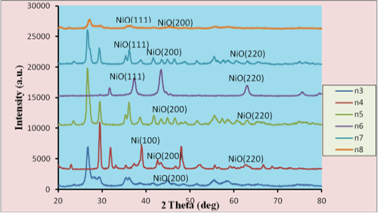
3.8.1. XRD Test
XRD test, which performed graphs considered the most important test and the first test in which the nanomaterial is tested. . And the graph in Figure 9 shows the presence of nanomaterial 37.02 (111), 44.6 (200)), 62.5 (220) in each of the NT6 and NT4 materials, and the results match the graph of the first method and the second method with international measurements of the Miller coefficient and the Theta degree 2.
Figure 9. Graph of N4, N6.
3.8.2. SEM Examination
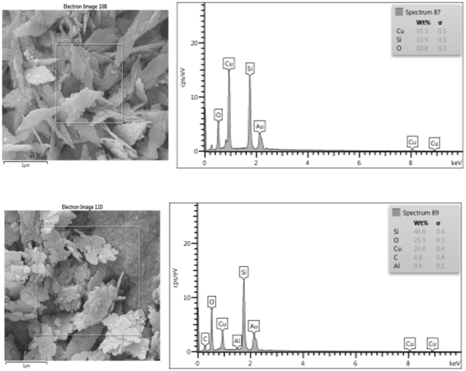
Figure 10. The graph showing the SEM results for the sample NT4 NT6.
It is noted from the Figure 10 N4 that the molecule shells or semi-shells smaller than 10 nm in size and N6, an figure that shows the molecule with a size of 20nm. SEM examination represents the details of the molecule and this examination of the sample may differ from one period to another for the same sample and the reason is due to many variables such as room temperature, storage period of the prepared material, preparation method, light, ambient air, humidity, solution components, sample weight as taking more than one examination of the sample does not give the same results at the same time .
3.8.3. TEM Examination
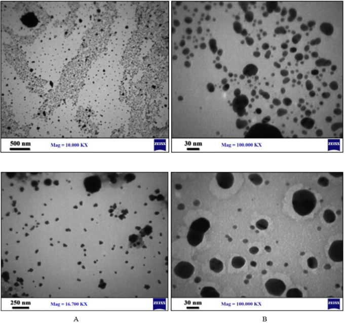
Figure 11. TEM of NT4, NT6.
It was noted from Figure 11, NT4 the largest scale of Figure11, NT4A was 500nm, and in Figure11, NT4B, which was of 30nm, are observed, exceeding sizes ranging from 0.5-30 nm. Figure 11 NT6, Imagre11, NT6A was 250nm, bodies in Figure11, NT6B, which was a drawing scale of 30nm, different sizes of nanoparticles are observed, not exceeding sizes ranging from 0.5 nm. These are particles called quantum dots according to the source .
3.8.4. UV Examination
Figure 12. UV, energy gap and absorbance of nickel oxide solution NT4, NT6.
It is noted from Figure 12, A NT4, the absorbance the opposite of the transmittance. If the transmittance value is low, the transmittance is high and vice versa. As for Graph B, it drawing value is 3.5 ev, which is a semiconductor material and it achieves the oxide of the nanomaterial. If the energy gap value is very large within the limits of the drawing, the material is an insulator such as glass and wood. It is noted from Figure 12, B UV absorbance and energy gap of nickel oxide solution with saffron extract, NT6 transmittance low absorbance in the range 236-800nm, which is very light, attributed the deviation the absorbance which for the NT6 sample, the energy gap value is 2.2 ev, and it achieves the oxide of the nanomaterial. .
3.8.5. FT-ir Examination
The material cannot be diagnosed or identified by the graph, but the energy of the active bonds absorbs heat and expands from the rays or heat emitted from the light. The movement of the vibrating materials is in six types of movements: sideways, forward, backward, upward and downward. These vibrating movements are called the frequency of the material F, and with the time factor they are called with a certain constant of the device FT, which is a sequence to convert time to space. The transformation of time indicates in the examination a function of the reciprocal of the wavelength, which is one to cm.
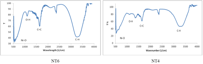
Figure 13. FT-IR NT4, NT6.
It is noted from the graphic Figure 13, A FT-IR of the nano-solutions of NT6, NT4, the sites of the metal oxide bonds Ni-O are at 500-600 and the wavelength 1210 is noted to be at the level of the C-O bond, 2400 may be O-H bond, 3300 N-H bond. [26].
3.8.6. Zeta Potential Test
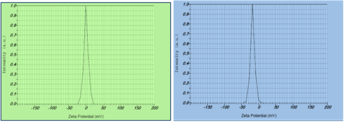
In Table 4 that there is a positive repulsive value, which is an indication of positive charges, while the negative values are the presence of negative repulsive. N4, which is the nano-solutions prepared by the first method, had a Zeta Potential graph value of 0.9 was negative were low, indicating that no clumping occurred in the nano-material. As for the nano-solution prepared by the second method, N6, its graph value in the test was -17.8 mv. negative were low, Figure 14 indicating that no clumping occurred in the nano-material .
Table 4. ZP results for the nano samples and the samples of two integrated nanomaterials.

Peak No.

Zeta Potential

Electrophoretic Mobility

N4

0.9 mv

0.000007 cm2/vs

N6

-17.8 mv

-0.000138 cm2/vs

Table 5. The rates of diameters of the bacterial growth inhibition zones as a result of the effect of nanoparticles.

ماء مقطر

Staphylococcus aureusتركيز. M%

Pseudomonas aerogenos تركيز. M%

0,0

0,1

0,01

0,05

0,07

0,1

0,01

0,05

0,07

ت

0

36

0

0

0

0

0

0

0

1

0

39

0

0

0

0

0

0

0

2

0

46

0

0

0

0

0

0

0

3

0

38

1

2

4

27

0

0

0

4

0

0

0

0

0

0

0

0

0

5

0

48

0

6

7

45

0

5

6

10

0

45

3

5

8

44

4

5

6

11

0

41

3

5

7

30

3

4

7

12

0

42

0

1

2

33

0

2

5

13

0

46

0

0

0

23

0

0

0

14

Figure 14. The graphs of the results of the zeta potential test for the NT4, NT6 nano samples.
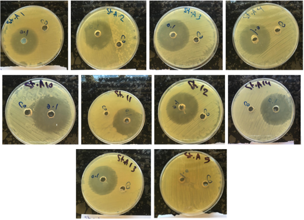
Figure 15. The diameters of inhibition of Staph. aureus growth using extracts of mixing two nanomaterials at ratios of 0.1 M%. Bacterial growth was observed in the solutions of mixing materials No. (1, 2, 3, 4, 5), and solutions (10, 11, 12, 13, 14).
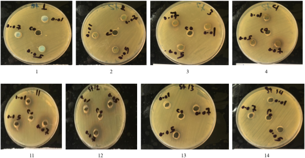
Figure 16. The diameters of inhibition of Staph. aureus growth using extracts of mixing two nanomaterials at ratios of (0.01, 0.05, 0.07) M%. Bacterial growth was observed in solutions No. (1, 2, 3, 4, 6), solutions of mixing the materials.
3.9. Nanological Examinations of Solutions in Which Two Nanomaterials Were Mixed
The examinations were conducted according to the method of .
3.9.1. XRD the Examination
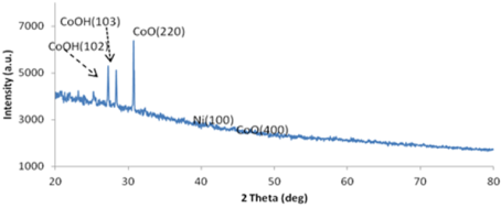
XRD The Examination Results were shown Specifications of Braak Angle in the Corresponding Graphs. The Specifications are Adopted. The Compound, Cobalt Oxide, is the Dominant one.
Figure 17. XRD examination CoO, NiO.
It was noted from the graph Figure 17 that the cobalt compound is dominant with the presence of the nickel compound and the appearance of two peaks of CoOH with Miller coefficient (102) and (103) at 2 Theta deg)) .
3.9.2. SEM Examination
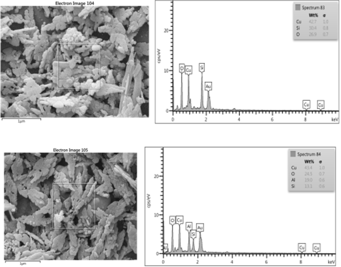
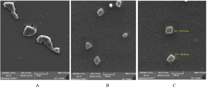
SEM examination showed the results as shown in the Figure 18, A, representing a material flower 200nm and measuring 50-90nm, Figure 18, B is a large Figure with dimensions of 10 µm and it is a flower. Figure 18, C shows measurements of different sides of the nanoparticle. The multiple shapes in the Figure may be attributed to nickel oxide, cobalt or other materials. SEM examination represents the details of the particle and this examination of the sample may differ from one period to another for the same sample. This is due to many variables such as room temperature, storage period of the prepared material, preparation method, light, ambient air, humidity, solution components, and sample weight. Taking more than one examination of the sample does not give the same results at the same time.
Figure 18. SEM examination result of the solution of the two nanomaterials.
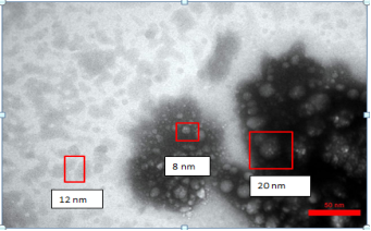
Figure 18, A shows measurement 50-90 nm smaller than 100 nm. As for Figure18, B, it is a large Figure with dimensions of 10 µm, and they are floral shapes of the nanoparticles, while Figure18, C shows measurements of different sides of the nanoparticle and the multiplicity of shapes in the Figure may be attributed to the fact that these particles indicate one of the materials, nickel oxide, cobalt or other materials.
3.9.3. TEM Examination
The results showed that through was fixed 50 nm was have that their size ranges between 5-12 nm, nanoparticles known worldwide are called quantum dots, according to the source (Poole, 2003), as shown in Figure 19.
Figure 19. TEM examination result of the solution of the two nanomaterial.
3.9.4. FT-ir Examination
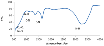
Figure 20. The FT-ir test (Quilt nitrate with aloe vera extract with nickel nitrate solution with saffron extract.
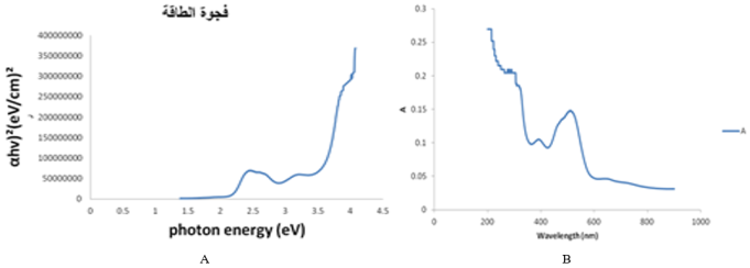
Figure 20 the locations of the metal oxide bonds Co-O and Ni-O are at 532, 1315 C-N bonds, 1600 C-N bonds carbon bonds, 3200 N-H bonds, 1080.
3.9.5. UV Examination
UV examination showed the results through the graph. Through the results that appeared, it was observed that the energy gap. As for the results through the examination, the energy gap energy 2.0ev, while the absorbance shows the formation of the plasma layer and at the wavelength 200-400 nm the x-axis wavelength (nm) This is evidence of the formation of the nanomaterial Figure 21.
Figure 21. UV is test CoO and NiO.
3.9.6. Zeta Potential
The results showed, as noted from the graph, there is a positive value of the repulsive, which is evidence of positive charges, while the negative values It is the presence of negatively charged particles. The value of the graph Zeta Potential is -27.5 mv, which is a low value that does not cause agglomeration. This applies to the results of the values of (coil nitrate with Aloe vera extract with nickel nitrate solution with saffron extract).
Table 6. The ZP results for the nano samples and the samples of two composite nanomaterials.

Peak No.

Zeta Potential

Electrophoretic Mobility

11

-36.7 mv

-0.000494 cm2/ vs

Where it was -36.7 mv, which are very low mv values, meaning that no clumping occurs in the nanomaterial as in Figure 22 This is consistent with what the researcher has reached.
Figure 22. Zeta potential.
4. MTT Toxicity Measurement
MTT toxicity measurement, the toxic substance live cells.
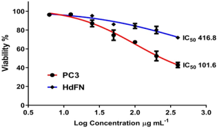
Figure 23. A result of MTT toxicity.

The IC50 ratio for killing 50% of cells for cancerous PC3 cells was observed to be 416.8, while its value for normal HdFN cells was 101.6 Table 7.
Table 7. The amount of SD values toxicity measurement MTT. The PC3, IC50 and HdFN. The amount of SD values toxicity measurement MTT. The PC3, IC50 and HdFN. The amount of SD values toxicity measurement MTT. The PC3, IC50 and HdFN.

Concen.

PC3

HdFN

Mean

SD

Mean

SD

400.00

42.82

2.77

3.00

71.95

0.81

200.00

52.43

5.12

3.00

80.03

3.88

100.00

67.25

1.45

3.00

84.34

2.66

50.00

74.54

5.42

3.00

86.07

1.92

25.00

87.08

3.51

3.00

95.22

0.82

12.50

96.76

1.14

3.00

95.95

1.03

6.25

96.30

0.81

3.00

95.95

0.20

5. Conclusions
1) The extract of cobalt nitrate with aloe vera is the most efficient plant extracts combined with nanomaterials in inhibiting the isolates of Staph. aureus and Ps. aeruginosa bacteria.
2) The results of the MTT cytotoxicity assay showed that the solutions prepared to treat infections caused by both Staph. aureus and Ps. aeruginosa were non-toxic solutions and highly effective when diluted to low concentrations.
6. Recommendations
Detection and investigation of phenotypic changes in the walls of bacterial cells exposed to the nanoparticles under study using transmission electron microscopy and X-ray diffract meter.
Abbreviations

FT-ir

Fourier Transform Infrared Spectroscopy

TEM

Transmission Electron Microscope

MTT

metabolic Dye [3-(4,5 Dimethyl Thiazol-2-yl)- 2, 5-] Diphenylterazolium$Bromide

MIC

Minimum Inhibitory Concentration

MBC

Minimum Bactericidal Concentration

UV

UV–visible Analysis

XRD

X-ray Diffraction

ZP

Zeta Potential

Author Contributions
Asmaa Ahmed Hatem Sultan: Conceptualization, Data curation, Formal Analysis, Funding acquisition, Investigation, Methodology, Project administration, Resources, Software, Visualization, Writing – original draft, Writing – review & editing
Conflicts of Interest
The authors declare no conflicts of interest.
References
[1] Cirnski, K., Coetzee, J., Herrmann, J., & Müller, R. Metabolic Profiling to Determine Bactericidal or Bacteriostatic Effects of New Natural Products using Isothermal Microcalorimetry. (2020).
[2] Kamel, Hassan Majeed. The effect of cumin fruit extracts and black pepper seeds in controlling food-source bacteria Master’s thesis, College of Education, Al-Qadisiyah University. (2018).
[3] Garcia-Espinoza, J. A., Aguilar-Aragon, V. B., Ortiz-Villalobos, E. H., Garcia-Manzano, R. A., & Antonio, B. A. Burns: definition, classification, pathophysiology and initial approach. Gen Med, (2020). 5(5), 1-5.
[4] Cerceo, E., Deitelzweig, S. B., Sherman, B. M., & Amin, A. N. Multidrug- resistant gram-negative bacterial infections in the hospital setting: overview, implications for clinical practice, and emerging treatment options. Microbial Drug Resistance, 22(5), (2016). 412-431.
[5] Ghazi, Riyam Thamer * Israa Ghanem Al-Samak. The synergistic effect of zinc nanoparticles and the antibiotic Erythromycin on methicillin-resistant Staphylococcus aureus bacteria isolated from different infections. Department of Life Sciences / College of Science / University of Mosul. (2020).
[6] Khazal, Laila Asi, Najdat Bahjat Mahdi 1. Study of the efficiency of some antibiotics of different origins on S. aureus bacteria isolated from different clinical sources. University of Kirkuk / College of General Studies / Department of Life Sciences (2015).
[7] Al-Rubaie, Abeer Fawzi Murad. Survey of the prevailing bacterial spores in Babylon Governorate, Master’s thesis, College of Science, University of Babylon. (2000).
[8] Nakagawa, S., Hillebrand, G. G., & Nunez, G. Rosmarinus officinalis l.(rosemary) extracts containing carnosic acid and carnosol are potent quorum sensing inhibitors of staphylococcus aureus virulence. Antsibiotics, (2020). 9(4), 149.
[9] Okuma, K., Iwakawa, K., Turnidge, J. D., Grubb, W. B., Bell, J. M., O'Brien, F. G., ... & Hiramatsu, K. Dissemination of new methicillin-resistant Staphylococcus aureus clones in the community. Journal of clinical microbiology, (2002). 40(11), 4289-4294.
[10] Foster, T. J. Immune evasion by staphylococci. Nature reviews microbiology, (2005). 3(12), 948-958.
[11] Weidenmaier, C., Kokai-Kun, J. F., Kristian, S. A., Chanturiya, T., Kalbacher, H., Gross, M., ... & Peschel, A. Role of teichoic acids in Staphylococcus aureus nasal colonization, a major risk factor in nosocomial infections. Nature medicine, (2004). 10(3), 243-245.
[12] Radovanovic, M., Petrovic, M., Hanna, R. D., Nordstrom, C. W., Calvin, A. D., Barsoum, M. K., ... & Dumic, I. Clinical Presentation and Management of Methicillin-Resistant Staphylococcus aureus Pericarditis—Systematic Review. Journal of Cardiovascular Development and Disease, (2022). 9(4), 103.
[13] Dinges, M. M., Orwin, P. M., & Schlievert, P. M. Exotoxins of Staphylococcus aureus. Clinical microbiology reviews, (2000). 13(1), 16-34. ‏
[14] Yousefi, M., Farshidi, M., Sani, M. A., Payahoo, L., & Ehsani, A. Investigation of the microbial quality and the occurrence of Shiga-like-toxin-producing Escherichia coli and methicillin resistant Staphylococcus aureus in traditional cheeses produced in northwest of Iran. Nutrition & Food Science. (2018).
[15] AL-Hilli, F. A. Study of Antimicrobial effect of leaves extract from Callistemoncitrinuson Pseudomonas aeruginosa isolated from patients (Doctoral dissertation, M. Sc. Thesis, Coll. Sci., Univ. AL-Mustansiriya). (2000).
[16] Payam, A. F., Biglarbeigi, P., Morelli, A., Lemoine, P., McLaughlin, J., & Finlay, D. Data acquisition and imaging using wavelet transform: a new path for high speed transient force microscopy. Nanoscale Advances, (2021). 3(2), 383-398.
[17] Xavier, D. E., Picão, R. C., Girardello, R., Fehlberg, L. C., & Gales, A. C. Efflux pumps expression and its association with porin down-regulation and β-lactamase production among Pseudomonas aeruginosa causing bloodstream infections in Brazil. BMC microbiology, (2010). 10(1), 1-7.
[18] Douglas, M. W., Mulholland, K., Denyer, V., & Gottlieb, T. Multi-drug resistant Pseudomonas aeruginosa outbreak in a burns (2001).
[19] Diggle, S. P., & Whiteley, M. Microbe Profile: Pseudomonas aeruginosa: opportunistic pathogen and lab rat. Microbiology, (2020). 166(1), 30.
[20] Nikaido, H. Multidrug resistance in bacteria. Annual review of biochemistry, (2009). 78, 119-146.
[21] Chanishvili, N., Chanishvili, T., Tediashvili, M., & Barrow, P. A. Phages and their application against drug‐resistant bacteria. Journal of Chemical Technology & Biotechnology, (2001).76(7), 689-699.
[22] (22). Langer, P. Mit schnellem Griff zu den Fakten: Taschenbuch Chemische Substanzen. (2002). Von A. Willmes. Harri Deutsch, Frankfurt aM 2001. 1195 Seiten, brosch. 29, 95 Euro, ISBN: 3‐8171‐1662‐4.
[23] Sun, S. S., Chen, Y., Yang, J., Tian, T. F., Deng, H. X., Li, W. H., ... & Alici, G. The development of an adaptive tuned magnetorheological elastomer absorber working in squeeze mode. Smart Materials and Structures, (2014). 23(7), 075009.
[24] Eftekhar, F., Rastegar, M., Golalipoor, M., & Mansoursamaei, N. Detection of Extended Spectrum B-Lactamases in Urinary Isolates of Klebsiella pneumoniae in Relation to blaSHV, blaTEM and blaCTX-M gene carriage. Iranian journal of public health, (2012). 41(3), 127.
[25] Enders, A., North, N., Fensore, C., Velez-Alvarez, J., & Allen, H. Functional group identification for FTIR spectra using Figure-based machine learning models. (2021).
[26] Malvern Instruments, L. Zeta Potential: An Introduction in 30 Minutes. Malvern Instruments. Report nr mrk, (2012). 654, 6.
[27] Musa, M. M. A. Antimicrobial activity of Aloe Barbadensis millar (Aloe Vera) against Pseudomonas aeruginosa isolates in Khartoum state, Sudan (Doctoral dissertation, Sudan University of Science & Technology). (2019).
[28] Srivastava, N., & Srivastava, P. C. Realizing NiO nanocrystals from a simple chemical method. Bulletin of Materials Science, (2010). 33(6), 653-656.
[29] Pal, S. L., Jana, U., Manna, P. K., Mohanta, G. P., & Manavalan, R. Nanoparticle: An overview of preparation and characterization. Journal of applied pharmaceutical science, (2011).1(6), 228-234.
[30] Jalill, R. D. A., Jawad, M. M. H. M., & Abd, A. N. Plants extracts as green synthesis of zirconium oxide nanoparticles. J. Genet. Environ. Res. Conserv, (2017). ‏‏ 5(1), 6-23.
[31] Jalill, A., Raghad, D. H., Nuaman, R. S., & Abd, A. N. Biological synthesis of Titanium Dioxide nanoparticles by Curcuma longa plant extract and study its biological properties. World Scientific News, (2016). 49(2), 204-222.
[32] Poole Jr, C. P., & Owens, F. J. Introduction to nanotechnology. John Wiley & Sons. (2003).
[33] Abd, A., Ali, R., & Hussein, A. Fabrication and characterization of nickel oxide nanoparticles/silicon heterojunction. J Multidiscip Eng Sci Stud, (2016). 2, 434-40.
[34] Hashim, A. A. (Ed.). Smart nanoparticles technology. BoD–Books on Demand. (2012).
[35] Abdel-Aal, S. K., Beskrovnyi, A. I., Ionov, A. M., Mozhchil, R. N., & Abdel-Rahman, A. S. Structure Investigation by Neutron Diffraction and X‐Ray Diffraction of Graphene Nanocomposite CuO–rGO Prepared by Low‐Cost Method. physica status solidi (a), (2021). 2100138.
[36] Kalyani, R., & Gurunathan, K. Intercalated network of graphene oxide (GO)–CuO–polythiophene (PTh) hybrid nanocomposite for photocatalytic applications. Journal of Materials Science: Materials in Electronics, (2016). 27(10), 10634-10641.
[37] El Bachiri, A., Soussi, L., Karzazi, O., Louardi, A., Rmili, A., Erguig, H., & El Idrissi, B. Electrochromic and photoluminescence properties of cobalt oxide thin films prepared by spray pyrolysis. Spectroscopy Letters, (2019) 52(1), 66-73.
Cite This Article
  • APA Style

    Sultan, A. A. H. (2026). Evaluation of the Inhibitory Effect and Cytotoxicity of Some Nanoparticles Against Bacterial Isolates from Burn Infections. International Journal of Clinical and Experimental Medical Sciences, 12(3), 39-56. https://doi.org/10.11648/j.ijcems.20261203.11

    Copy | Download

    ACS Style

    Sultan, A. A. H. Evaluation of the Inhibitory Effect and Cytotoxicity of Some Nanoparticles Against Bacterial Isolates from Burn Infections. Int. J. Clin. Exp. Med. Sci. 2026, 12(3), 39-56. doi: 10.11648/j.ijcems.20261203.11

    Copy | Download

    AMA Style

    Sultan AAH. Evaluation of the Inhibitory Effect and Cytotoxicity of Some Nanoparticles Against Bacterial Isolates from Burn Infections. Int J Clin Exp Med Sci. 2026;12(3):39-56. doi: 10.11648/j.ijcems.20261203.11

    Copy | Download

  • @article{10.11648/j.ijcems.20261203.11,
      author = {Asmaa Ahmed Hatem Sultan},
      title = {Evaluation of the Inhibitory Effect and Cytotoxicity of Some Nanoparticles Against Bacterial Isolates from Burn Infections},
      journal = {International Journal of Clinical and Experimental Medical Sciences},
      volume = {12},
      number = {3},
      pages = {39-56},
      doi = {10.11648/j.ijcems.20261203.11},
      url = {https://doi.org/10.11648/j.ijcems.20261203.11},
      eprint = {https://article.sciencepublishinggroup.com/pdf/10.11648.j.ijcems.20261203.11},
      abstract = {150 swabs were taken from different burns of patients admitted to Medical City Hospital and Al-Kindi Teaching Hospital. 128 swabs showed morphological examinations. The isolates were confirmed at the species level using Vitek2 compact system for skin diseases. The isolates under study were tested for sensitivity using the drill diffusion method. Nanoparticles were prepared by two methods, chemical synthesis and green synthesis, Aloe vera and aqueous extract of saffron (Crocus sativus L.). Bacteria were isolated by 60 isolates of Pseudomonas aeruginosa, (46.87%), followed by Staphylococcus aureus, with 38 isolates (12.28%), Klebsiella spp. with 20 isolates (15.62%), and the rest of the isolates were Escherichia coli, Enterobacter cloacae, Acinetobacter baumannii, Stenotrophomonas maltophilia, with three isolates for each type, i.e. (1.56%). S. aureus and Ps. aeruginosa were the most resistant bacteria to antibiotics, and it was found that substance nanoparticle prepared by Artificial roads nanoparticle. The percentage of Staph. aureus bacteria was (28.12%), while Ps. aeruginosa bacteria was (46.87%). The solutions were at concentrations of (100, 75, 50, 25) M%. It is noted that the highest inhibition concentration was (100) M%. As for the is Staph. aureus and Ps. aeruginosa bacteria isolates zone (48, 45) mm, respectively. It was found that the saffron of (45) mm in inhibiting the isolates of Gram-positive bacteria Staph. aureus. The success of Staph. aureus with nickel oxide with saffron at a rate of MIC. Invivo test animals, and the MBC the conducted two methods tests were, ZETA POTENTIAL. Nanomaterials with a smaller size of 5-12nm called quantum dot were observed, and no agglomeration occurred in them. It was shown that the MTT toxicity assay for the solutions of the IC50 ratio for cancerous PC3, while for cellsand the substance is not toxic to the solution for treating S. aureus bacteria, and Ps. aeruginosa.},
     year = {2026}
    }
    

    Copy | Download

  • TY  - JOUR
    T1  - Evaluation of the Inhibitory Effect and Cytotoxicity of Some Nanoparticles Against Bacterial Isolates from Burn Infections
    AU  - Asmaa Ahmed Hatem Sultan
    Y1  - 2026/08/17
    PY  - 2026
    N1  - https://doi.org/10.11648/j.ijcems.20261203.11
    DO  - 10.11648/j.ijcems.20261203.11
    T2  - International Journal of Clinical and Experimental Medical Sciences
    JF  - International Journal of Clinical and Experimental Medical Sciences
    JO  - International Journal of Clinical and Experimental Medical Sciences
    SP  - 39
    EP  - 56
    PB  - Science Publishing Group
    SN  - 2469-8032
    UR  - https://doi.org/10.11648/j.ijcems.20261203.11
    AB  - 150 swabs were taken from different burns of patients admitted to Medical City Hospital and Al-Kindi Teaching Hospital. 128 swabs showed morphological examinations. The isolates were confirmed at the species level using Vitek2 compact system for skin diseases. The isolates under study were tested for sensitivity using the drill diffusion method. Nanoparticles were prepared by two methods, chemical synthesis and green synthesis, Aloe vera and aqueous extract of saffron (Crocus sativus L.). Bacteria were isolated by 60 isolates of Pseudomonas aeruginosa, (46.87%), followed by Staphylococcus aureus, with 38 isolates (12.28%), Klebsiella spp. with 20 isolates (15.62%), and the rest of the isolates were Escherichia coli, Enterobacter cloacae, Acinetobacter baumannii, Stenotrophomonas maltophilia, with three isolates for each type, i.e. (1.56%). S. aureus and Ps. aeruginosa were the most resistant bacteria to antibiotics, and it was found that substance nanoparticle prepared by Artificial roads nanoparticle. The percentage of Staph. aureus bacteria was (28.12%), while Ps. aeruginosa bacteria was (46.87%). The solutions were at concentrations of (100, 75, 50, 25) M%. It is noted that the highest inhibition concentration was (100) M%. As for the is Staph. aureus and Ps. aeruginosa bacteria isolates zone (48, 45) mm, respectively. It was found that the saffron of (45) mm in inhibiting the isolates of Gram-positive bacteria Staph. aureus. The success of Staph. aureus with nickel oxide with saffron at a rate of MIC. Invivo test animals, and the MBC the conducted two methods tests were, ZETA POTENTIAL. Nanomaterials with a smaller size of 5-12nm called quantum dot were observed, and no agglomeration occurred in them. It was shown that the MTT toxicity assay for the solutions of the IC50 ratio for cancerous PC3, while for cellsand the substance is not toxic to the solution for treating S. aureus bacteria, and Ps. aeruginosa.
    VL  - 12
    IS  - 3
    ER  - 

    Copy | Download

Author Information
  • Abstract
  • Keywords
  • Document Sections

    1. 1. Introduction
    2. 2. Materials and Method
    3. 3. Results and Discussion
    4. 4. MTT Toxicity Measurement
    5. 5. Conclusions
    6. 6. Recommendations
    Show Full Outline
  • Abbreviations
  • Author Contributions
  • Conflicts of Interest
  • References
  • Cite This Article
  • Author Information