Research Article | | Peer-Reviewed

Corticosteroid-Induced Candida Albicans Colonization of an Intragastric Balloon: A Case Report and Literature Review

Received: 17 July 2026     Accepted: 4 August 2026     Published: 20 August 2026
Views:       Downloads:
Abstract

Background: Intragastric balloons (IGBs) are temporary endoscopic devices used for weight management in patients with obesity; while bacterial colonization of IGBs is well recognized, fungal infection remains rarely reported. Case Presentation: We describe a 24-year-old male who developed candidal infection of an intragastric balloon after completing a 14-day course of dexamethasone (8 mg daily), equivalent to approximately 53 mg of prednisone daily, prescribed for presumed allergic rhinitis. Six weeks after balloon placement for obesity management (body mass index [BMI]: 37.2 kg/m²), he presented with epigastric discomfort, nausea, and intermittent vomiting. Upper gastrointestinal endoscopy revealed extensive white-cream fungal plaques covering the balloon surface with mild surrounding mucosal erythema. Microbiological analysis of gastric aspirate confirmed Candida albicans (>10⁵ colony-forming units [CFU]/mL) on culture, with positive potassium hydroxide (KOH) preparation and Periodic Acid-Schiff (PAS) staining. The balloon was removed endoscopically and the patient was treated with fluconazole (400 mg loading dose, then 200 mg daily for 14 days), with complete symptom resolution within five days and eradication confirmed on repeat endoscopy and culture at four weeks. Conclusion: This case illustrates that even short-term corticosteroid therapy can predispose patients with an indwelling IGB to opportunistic fungal colonization. Clinicians should maintain a high index of suspicion for fungal infection in IGB patients who develop new gastrointestinal symptoms during or after immunosuppressive therapy, and early endoscopic evaluation, balloon removal, and antifungal therapy achieve favorable outcomes.

Published in International Journal of Gastroenterology (Volume 10, Issue 2)
DOI 10.11648/j.ijg.20261002.12
Page(s) 74-79
Creative Commons

This is an Open Access article, distributed under the terms of the Creative Commons Attribution 4.0 International License (http://creativecommons.org/licenses/by/4.0/), which permits unrestricted use, distribution and reproduction in any medium or format, provided the original work is properly cited.

Copyright

Copyright © The Author(s), 2026. Published by Science Publishing Group

Keywords

Intragastric Balloon, Fungal Infection, Dexamethasone, Immunosuppression, Obesity Management

1. Introduction
Intragastric balloons have emerged as a minimally invasive option for weight management in patients with obesity who have failed conservative measures. These devices are typically maintained for 6–12 months and work through gastric volume restriction and early satiety induction. While generally safe, complications including nausea, vomiting, gastric ulceration, and balloon deflation have been documented. Microbial colonization of IGBs has been reported, predominantly involving bacterial species. However, fungal infections remain exceedingly rare, with limited literature on their occurrence and management.
Corticosteroids are known to suppress cell-mediated immunity, increasing susceptibility to opportunistic infections, particularly fungal pathogens. We present a unique case of fungal colonization of an intragastric balloon in a young patient following short-term dexamethasone therapy.
2. Case Scenario
2.1. Initial Presentation
A 24-year-old male presented to the gastroenterology clinic with a three-day history of epigastric discomfort, nausea, and intermittent vomiting. He reported no hematemesis, melena, or fever. The patient had undergone intragastric balloon placement six weeks prior for obesity management (BMI: 37.2 kg/m2).
2.2. Medical History
1) Obesity: Long-standing, failed multiple dietary interventions.
2) Intragastric Balloon: Fluid-filled saline balloon, placed 6 weeks prior to presentation.
3) Recent Medication: Dexamethasone 8 mg daily for 14 days, prescribed by another physician for presumed allergic rhinitis.
4) No history of: Diabetes mellitus, immunodeficiency, chronic infections, or previous fungal infections.
2.3. Medication History
Two weeks prior to symptom onset, the patient was prescribed dexamethasone 8 mg daily by an otolaryngologist for management of persistent nasal symptoms initially thought to be allergic rhinitis. He completed a 14-day course, with the last dose taken three days before his current presentation.
2.4. Clinical Examination
1) Vital Signs: Temperature 37.1°C, BP 128/76 mmHg, HR 82 bpm.
2) General: Alert, not in acute distress.
3) Abdominal Examination: Mild epigastric tenderness, no guarding or rebound, normal bowel sounds.
4) Oral Examination: No evidence of oral thrush.
2.5. Endoscopic Findings
Upper gastrointestinal endoscopy was performed and revealed:
1) Intragastric balloon in situ, properly positioned in the gastric fundus.
2) Multiple white-cream colored patches and plaques adherent to the balloon surface.
3) Mild surrounding gastric mucosal erythema.
4) No ulceration or balloon deflation noted.
5) Gastric mucosa otherwise normal.
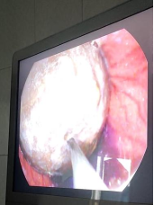
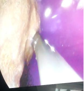
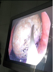
2.5.1. In Situ Endoscopic Findings (Figures 1–3)
The endoscopic examination revealed striking findings consistent with severe fungal colonization: the intragastric balloon was properly positioned within the gastric fundus, with extensive white to cream-colored plaques and patches covering significant portions of the balloon surface. Fungal colonies appeared as confluent areas interspersed with discrete smaller plaques, with irregular borders and varying thickness. The underlying balloon surface remained intact without evidence of deflation or rupture. Surrounding gastric mucosa demonstrated mild inflammatory changes with erythema, but no gastric ulceration, erosions, or bleeding was noted, and normal peristaltic activity was observed.
Figure 1. Endoscopic view of the intragastric balloon showing extensive white-cream fungal plaques with a smooth balloon contour; the endoscope tip is visible in the field.
Figure 2. Close-up endoscopic view demonstrating confluent white-yellowish fungal colonies with irregular borders and mild surrounding gastric mucosal erythema.
Figure 3. Endoscopic view highlighting the distribution of fungal colonization across the balloon surface, showing confluent and scattered plaque patterns typical of Candida infection.
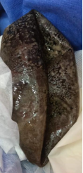
2.5.2. Post-Removal Macroscopic Examination (Figure 4)
The removed balloon demonstrated dramatic gross pathological changes: extensive dark brown to black discoloration covering large surface areas, with dense, tenacious fungal biofilm firmly adherent to the balloon material. The fungal colonies formed thick, leathery deposits that resisted mechanical removal, and surface texture was markedly altered from smooth to rough and irregular. The dramatic color change from the typical transparent/translucent balloon appearance to dark brown-black indicated heavy fungal burden and possible biofilm maturation. Balloon integrity was maintained despite severe colonization, with an estimated 60–70% of the total balloon surface area affected by visible fungal growth.
The contrast between the endoscopic appearance (white-cream plaques) and the post-removal appearance (dark brown-black deposits) likely represents different stages of fungal colonization and biofilm development, with the darker appearance indicating mature, densely organized fungal communities.
Figure 4. Gross specimen photograph of the removed intragastric balloon showing extensive dark brown-black discoloration with adherent fungal biofilm covering large portions of the surface.
2.6. Investigations
2.6.1. Laboratory Studies
1) Complete Blood Count: WBC 9,200/µL (normal differential).
2) C-Reactive Protein (CRP): 12 mg/L (mildly elevated).
3) Renal and liver function: Within normal limits.
4) HIV serology: Negative.
5) Fasting glucose: 92 mg/dL.
2.6.2. Microbiological Studies
1) Gastric aspirate collected during endoscopy.
2) Direct microscopic examination with KOH preparation: Positive for fungal elements (budding yeasts and pseudohyphae).
3) Fungal culture: Candida albicans isolated (>105 CFU/mL).
4) Bacterial cultures: Negative for pathogenic organisms.
5) Antifungal susceptibility testing: Sensitive to fluconazole, itraconazole, and amphotericin B.
2.6.3. Histopathology
1) Brush cytology from balloon surface showed numerous fungal organisms consistent with Candida species with characteristic budding yeast forms.
2) PAS (Periodic Acid-Schiff) stain: Positive, confirming fungal elements.
3) Balloon surface material examination: Dense fungal biofilm with mixed morphological forms (yeast and pseudohyphal elements).
4) No malignant cells identified.
2.7. Case Study Analysis
2.7.1. Diagnosis
Primary Diagnosis: Fungal infection (Candida albicans) of intragastric balloon.
Contributing Factor: Iatrogenic immunosuppression secondary to corticosteroid therapy.
2.7.2. Pathophysiology
The development of severe fungal colonization in this case can be attributed to multiple synergistic factors:
1) Corticosteroid-Induced Immunosuppression: Dexamethasone suppresses T-cell mediated immunity and impairs neutrophil function, creating favorable conditions for opportunistic fungal infections. Even short-term use (2 weeks) at moderate-to-high doses can significantly increase infection risk. The dose of 8 mg daily represents substantial immunosuppression, equivalent to approximately 53 mg of prednisone daily.
2) Foreign Body Presence and Biofilm Formation: The intragastric balloon serves as an ideal substrate for microbial adherence and complex biofilm formation. As demonstrated in Figure 4, the heavy dark brown-black discoloration represents mature, multilayered biofilm with extensive fungal communities that confer protection against host immune responses and increase resistance to antifungal agents.
3) Altered Gastric Environment: The presence of the balloon may alter gastric pH, motility, and mucosal integrity, potentially reducing local antimicrobial defenses and facilitating fungal colonization.
4) Candida Pathogenicity and Virulence Factors: Candida albicans possesses adhesins, biofilm-forming capability, morphological switching between yeast and hyphal forms, and protease/phospholipase production that enhance tissue invasion.
5) Progressive Colonization Pattern: The images reveal a continuum of fungal growth from early colonization (white-cream plaques seen endoscopically) to mature, organized biofilm (dark brown-black deposits on the removed specimen), indicating progressive fungal proliferation over time.
2.7.3. Management Strategy
Immediate Management:
1) Balloon Removal: Given the symptomatic infection and fungal colonization, early balloon removal was recommended and performed endoscopically without complications.
2) Antifungal Therapy: Fluconazole 400 mg loading dose, followed by 200 mg daily for 14 days, initiated immediately after balloon removal, in line with standard treatment guidelines for candidiasis.
3) Symptomatic Management: Proton pump inhibitor (omeprazole 40 mg daily) and antiemetics as needed.
Follow-Up Management:
1) Clinical reassessment at 1 week: Complete resolution of symptoms.
2) Repeat endoscopy at 4 weeks: Normal gastric mucosa, no residual infection.
3) Fungal culture from gastric aspirate: Negative.
4) Counseling regarding alternative weight management strategies.
2.7.4. Clinical Outcome
The patient experienced complete resolution of symptoms within 5 days of balloon removal and antifungal therapy initiation. Follow-up endoscopy confirmed eradication of the fungal infection with healing of gastric mucosa. The patient was subsequently enrolled in a comprehensive weight management program including dietary counseling, behavioral therapy, and supervised exercise.
3. Discussion
This case represents a rare complication of intragastric balloon therapy and highlights several important clinical considerations.
3.1. Incidence and Risk Factors
Fungal colonization of intragastric balloons is rarely reported in the literature, with most microbial complications involving bacterial organisms. Large device-safety analyses report major complication rates below 1.5% and mortality below 0.1% for IGBs overall, indicating that fungal colonization, though uncommon, represents a clinically important and likely underrecognized subset of these complications. Risk factors for fungal infection in IGB patients include:
1) Immunosuppressive therapy (corticosteroids, chemotherapy, biologics).
2) Diabetes mellitus with poor glycemic control.
3) Prolonged antibiotic use altering the gastric microbiome
4) Underlying immunodeficiency states
5) Proton pump inhibitor use (controversial).
3.2. Corticosteroid Effect
Even short-term corticosteroid therapy, as in this case, can significantly impair immune function. Dexamethasone at 8 mg daily for 14 days represents substantial immunosuppression, equivalent to approximately 53 mg of prednisone daily. This level of exposure is sufficient to increase opportunistic infection risk, particularly for fungal pathogens like Candida species. [7, 10, 11]
3.3. Clinical Implications
1) Screening: Patients with IGBs who require corticosteroid therapy should be counseled about infection risks and monitored closely.
2) Prophylaxis: In patients requiring prolonged immunosuppression, consideration should be given to antifungal prophylaxis or early balloon removal.
3) Early Recognition: New gastrointestinal symptoms in IGB patients with immunosuppression should prompt early endoscopic evaluation.
4) Management Approach: Balloon removal combined with appropriate antifungal therapy represents the definitive treatment for established fungal infection.
3.4. Literature Review
A review of the literature reveals very few published cases of fungal IGB infections. Most reports describe isolated cases of bacterial or fungal colonization, biofilm formation, or balloon hyperinflation attributed to gas-producing organisms, most commonly Candida species. This paucity of fungal infection reports may reflect the true rarity of the condition, underdiagnosis in asymptomatic patients, or limited routine endoscopic surveillance in IGB patients.
4. Conclusion
This case demonstrates that fungal infection of intragastric balloons, though rare, can occur in patients receiving immunosuppressive therapy, even with short-term corticosteroid use. The combination of a foreign body and compromised immunity creates favorable conditions for opportunistic fungal colonization. Clinicians should maintain heightened awareness of this potential complication, particularly in patients requiring corticosteroid therapy. Early recognition, prompt balloon removal, and appropriate antifungal treatment ensure favorable outcomes. This case underscores the importance of careful patient selection for IGB placement and ongoing surveillance for infectious complications in at-risk populations.
5. Recommendations
Based on this case, we propose the following preventive and clinical practice measures:
1) Conduct a thorough medical history assessment before IGB placement, including medication review.
2) Avoid IGB placement in patients requiring chronic immunosuppression.
3) Consider early balloon removal if significant immunosuppressive therapy becomes necessary.
4) Consider antifungal prophylaxis in high-risk patients.
5) Provide enhanced patient education regarding infection warning signs.
6) Maintain a high index of suspicion: new gastrointestinal symptoms in IGB patients with recent immunosuppression warrant prompt endoscopic evaluation, and management requires both balloon removal and systemic antifungal therapy.
Abbreviations

IGB

Intragastric Balloon

BMI

Body Mass Index

BP

Blood Pressure

HR

Heart Rate

CRP

C-Reactive Protein

WBC

White Blood Cell (count)

KOH

Potassium Hydroxide

PAS

Periodic Acid-Schiff

CFU

Colony-Forming Unit

HIV

Human Immunodeficiency Virus

Acknowledgments
Written informed consent was obtained from the patient for publication of this case report and accompanying images. The authors would like to thank the patient for providing consent for publication of this case report and the medical staff who assisted in patient care.
Author Contributions
Atteyat Aboelmaged Semeya: Conceptualization, Investigation, Methodology, Project administration, Supervision, Writing – original draft, Writing – review & editing
Walaa Elsaeed: Data curation, Investigation, Writing – original draft, Writing – review & editing
Mahmoud Ahmed Hassan: Investigation, Resources, Formal Analysis, Writing – review & editing
Shahenda Hossam: Investigation, Formal Analysis, Data curation, Writing – review & editing
Conflicts of Interest
The authors declare no conflicts of interest.
References
[1] Genco A, Cipriano M, Bacci V, et al. BioEnterics Intragastric Balloon (BIB): a short-term, double-blind, randomised, controlled, crossover study on weight reduction in morbidly obese patients. Int J Obes. 2006; 30(1): 129-133.
[2] Kumar N, Sahdala HN, Shaikh M, et al. Intragastric Balloons: Past, Present, and Future. Obes Surg. 2020; 30(2): 670-675.
[3] Stavrou G, Shrewsbury A, Kotzampassi K. Six intragastric balloons: Which to choose? World J Gastrointest Endosc. 2021; 13(8): 238-259.
[4] De Castro ML, Morales MJ, Del Campo V, et al. Efficacy, Safety, and Tolerance of Two Types of Intragastric Balloons Placed in Obese Subjects: A Double-Blind Comparative Study. Obes Surg. 2010; 20(12): 1642-1646.
[5] Pasha SF, Acosta RD, Chandrasekhara V, et al. The role of endoscopy in the bariatric surgery patient. Gastrointest Endosc. 2015; 81(5): 1063-1072.
[6] Pfaller MA, Diekema DJ. Epidemiology of invasive candidiasis: a persistent public health problem. Clin Microbiol Rev. 2007; 20(1): 133-163.
[7] Rhen T, Cidlowski JA. Antiinflammatory action of glucocorticoids—new mechanisms for old drugs. N Engl J Med. 2005; 353(16): 1711-1723.
[8] Prado A, Brito RO, Pereira ECA, Becker TCA, Negri M, Svidzinski TIE. The fungus Candida glabrata in intragastric balloon. Int J Case Rep Images. 2021; 12: 101246Z01AP2021.
[9] Guirgis M, Kumar P, Laurens J, Madhok B. Spontaneous Hyperinflation of Intragastric Balloons: A Systematic Review. Gastrointest Disord. 2025; 7(3): 55.
[10] Chastain DB, Spradlin M, Ahmad H, Henao-Martínez AF. Unintended Consequences: Risk of Opportunistic Infections Associated With Long-term Glucocorticoid Therapies in Adults. Clin Infect Dis. 2024; 78(4): e37-e56.
[11] Kim MF, et al. Glucocorticoids as a risk factor for infection and adverse outcomes in non-HIV and non-transplant patients with cryptococcal meningitis. Mycoses. 2024; 67: e13709.
[12] Kumar D, Rani A, Awani N. Molecular Determinants Involved in Candida albicans Biofilm Formation and Regulation. Mol Biotechnol. 2024; 66(7): 1640-1659.
[13] Idris I, Anyiam O. Intragastric balloons for obesity: critical review of device design, efficacy, tolerability, and unmet clinical needs. Diabetes Obes Metab. 2025; 27(Suppl 2): 20-34.
[14] Espinet-Coll E, Del Pozo-García AJ, Turró-Arau R, et al. Evaluating the Safety of the Intragastric Balloon: Spanish Multicenter Experience in 20,680 Cases and with 12 Different Balloon Models. Obes Surg. 2024; 34(8): 2766-2777.
[15] Goodman MC, Chang P, Minteer W, Nguyen D, Gopalkrishnan K, Phan J. Patient Complications and Device Issues Associated With FDA-Approved Intragastric Balloons Available in the USA: A MAUDE Database Study. Proc (Bayl Univ Med Cent). 2024; 38(1): 63-68.
[16] Edathodu Z, Khan SA, Peedikayil MC. Comparative Efficacy and Long-Term Outcomes of Intragastric Balloons for Obesity: A Systematic Review and Meta-Analysis. Cureus. 2025; 17(7): e88002.
[17] Pappas PG, Kauffman CA, Andes DR, et al. Clinical Practice Guideline for the Management of Candidiasis: 2016 Update by the Infectious Diseases Society of America. Clin Infect Dis. 2016; 62(4): e1-e50.
Cite This Article
  • APA Style

    Semeya, A. A., Elsaeed, W., Hassan, M. A., Hossam, S. (2026). Corticosteroid-Induced Candida Albicans Colonization of an Intragastric Balloon: A Case Report and Literature Review. International Journal of Gastroenterology, 10(2), 74-79. https://doi.org/10.11648/j.ijg.20261002.12

    Copy | Download

    ACS Style

    Semeya, A. A.; Elsaeed, W.; Hassan, M. A.; Hossam, S. Corticosteroid-Induced Candida Albicans Colonization of an Intragastric Balloon: A Case Report and Literature Review. Int. J. Gastroenterol. 2026, 10(2), 74-79. doi: 10.11648/j.ijg.20261002.12

    Copy | Download

    AMA Style

    Semeya AA, Elsaeed W, Hassan MA, Hossam S. Corticosteroid-Induced Candida Albicans Colonization of an Intragastric Balloon: A Case Report and Literature Review. Int J Gastroenterol. 2026;10(2):74-79. doi: 10.11648/j.ijg.20261002.12

    Copy | Download

  • @article{10.11648/j.ijg.20261002.12,
      author = {Atteyat Aboelmaged Semeya and Walaa Elsaeed and Mahmoud Ahmed Hassan and Shahenda Hossam},
      title = {Corticosteroid-Induced Candida Albicans Colonization of an Intragastric Balloon: A Case Report and Literature Review},
      journal = {International Journal of Gastroenterology},
      volume = {10},
      number = {2},
      pages = {74-79},
      doi = {10.11648/j.ijg.20261002.12},
      url = {https://doi.org/10.11648/j.ijg.20261002.12},
      eprint = {https://article.sciencepublishinggroup.com/pdf/10.11648.j.ijg.20261002.12},
      abstract = {Background: Intragastric balloons (IGBs) are temporary endoscopic devices used for weight management in patients with obesity; while bacterial colonization of IGBs is well recognized, fungal infection remains rarely reported. Case Presentation: We describe a 24-year-old male who developed candidal infection of an intragastric balloon after completing a 14-day course of dexamethasone (8 mg daily), equivalent to approximately 53 mg of prednisone daily, prescribed for presumed allergic rhinitis. Six weeks after balloon placement for obesity management (body mass index [BMI]: 37.2 kg/m²), he presented with epigastric discomfort, nausea, and intermittent vomiting. Upper gastrointestinal endoscopy revealed extensive white-cream fungal plaques covering the balloon surface with mild surrounding mucosal erythema. Microbiological analysis of gastric aspirate confirmed Candida albicans (>10⁵ colony-forming units [CFU]/mL) on culture, with positive potassium hydroxide (KOH) preparation and Periodic Acid-Schiff (PAS) staining. The balloon was removed endoscopically and the patient was treated with fluconazole (400 mg loading dose, then 200 mg daily for 14 days), with complete symptom resolution within five days and eradication confirmed on repeat endoscopy and culture at four weeks. Conclusion: This case illustrates that even short-term corticosteroid therapy can predispose patients with an indwelling IGB to opportunistic fungal colonization. Clinicians should maintain a high index of suspicion for fungal infection in IGB patients who develop new gastrointestinal symptoms during or after immunosuppressive therapy, and early endoscopic evaluation, balloon removal, and antifungal therapy achieve favorable outcomes.},
     year = {2026}
    }
    

    Copy | Download

  • TY  - JOUR
    T1  - Corticosteroid-Induced Candida Albicans Colonization of an Intragastric Balloon: A Case Report and Literature Review
    AU  - Atteyat Aboelmaged Semeya
    AU  - Walaa Elsaeed
    AU  - Mahmoud Ahmed Hassan
    AU  - Shahenda Hossam
    Y1  - 2026/08/20
    PY  - 2026
    N1  - https://doi.org/10.11648/j.ijg.20261002.12
    DO  - 10.11648/j.ijg.20261002.12
    T2  - International Journal of Gastroenterology
    JF  - International Journal of Gastroenterology
    JO  - International Journal of Gastroenterology
    SP  - 74
    EP  - 79
    PB  - Science Publishing Group
    SN  - 2640-169X
    UR  - https://doi.org/10.11648/j.ijg.20261002.12
    AB  - Background: Intragastric balloons (IGBs) are temporary endoscopic devices used for weight management in patients with obesity; while bacterial colonization of IGBs is well recognized, fungal infection remains rarely reported. Case Presentation: We describe a 24-year-old male who developed candidal infection of an intragastric balloon after completing a 14-day course of dexamethasone (8 mg daily), equivalent to approximately 53 mg of prednisone daily, prescribed for presumed allergic rhinitis. Six weeks after balloon placement for obesity management (body mass index [BMI]: 37.2 kg/m²), he presented with epigastric discomfort, nausea, and intermittent vomiting. Upper gastrointestinal endoscopy revealed extensive white-cream fungal plaques covering the balloon surface with mild surrounding mucosal erythema. Microbiological analysis of gastric aspirate confirmed Candida albicans (>10⁵ colony-forming units [CFU]/mL) on culture, with positive potassium hydroxide (KOH) preparation and Periodic Acid-Schiff (PAS) staining. The balloon was removed endoscopically and the patient was treated with fluconazole (400 mg loading dose, then 200 mg daily for 14 days), with complete symptom resolution within five days and eradication confirmed on repeat endoscopy and culture at four weeks. Conclusion: This case illustrates that even short-term corticosteroid therapy can predispose patients with an indwelling IGB to opportunistic fungal colonization. Clinicians should maintain a high index of suspicion for fungal infection in IGB patients who develop new gastrointestinal symptoms during or after immunosuppressive therapy, and early endoscopic evaluation, balloon removal, and antifungal therapy achieve favorable outcomes.
    VL  - 10
    IS  - 2
    ER  - 

    Copy | Download

Author Information
  • Department of Hepatogastroenterology, Benha Teaching Hospital, Benha, Egypt
  • Department of Hepatogastroenterology, Benha Teaching Hospital, Benha, Egypt

  • Department of Hepatogastroenterology, Benha Teaching Hospital, Benha, Egypt

  • Department of Hepatogastroenterology, Benha Teaching Hospital, Benha, Egypt

  • Abstract
  • Keywords
  • Document Sections

    1. 1. Introduction
    2. 2. Case Scenario
    3. 3. Discussion
    4. 4. Conclusion
    5. 5. Recommendations
    Show Full Outline
  • Abbreviations
  • Acknowledgments
  • Author Contributions
  • Conflicts of Interest
  • References
  • Cite This Article
  • Author Information