Review Article | | Peer-Reviewed

A Review of CT-Guided Lung Biopsy: Procedure Stages, Complications, and Risk Mitigation

Received: 21 December 2025     Accepted: 26 February 2026     Published: 3 July 2026
Views:       Downloads:
Abstract

The modern method of diagnosis of lung lesions, biopsy guided by CT is widely used in clinical practice. Especially this method is used frequently in oncological hospitals. In our study we analyzed data of patients of the oncology institute, who underwent a lung biopsy using a biopsy guided by CTand complications associated with this diagnostic procedure. The aim of our study was to objectively evaluate the types of complications in patients after the procedure, to compare them with data from the preliminary examination in order to reduce or avoid them in the future. Among all patients who underwent this procedure (89), the number of serious complications during this period of 1.5 years was: pneumothorax 5.6%, hemorrhage 1.1%, hemoptisis - upto 3.3%. In our opinion, the elevation of the risks is influenced by long smoking history (clinical data, coagulation disorders). We discuss in detail the tactics of management and observation of patients after the procedure, taking into account the patient's medical history and complications during the procedure. Careful preliminary preparation of the patients before the procedure helps to reduce the risk of complications.

Published in International Journal of Pharmacy and Chemistry (Volume 12, Issue 2)
DOI 10.11648/j.ijpc.20261202.11
Page(s) 19-23
Creative Commons

This is an Open Access article, distributed under the terms of the Creative Commons Attribution 4.0 International License (http://creativecommons.org/licenses/by/4.0/), which permits unrestricted use, distribution and reproduction in any medium or format, provided the original work is properly cited.

Copyright

Copyright © The Author(s), 2026. Published by Science Publishing Group

Keywords

CT, Lung Biopsy Guided by CT, Lung Injury, Pneumothorax, Pulmonary Hemorrhage/Hemoptysis

1. Introduction
Currently, the modern methods of diagnostic, such as CT, PET-CT, MRI are widely used to perform a diagnosis in oncology. In the differential diagnosis of the pathology of the organs of the chest, the CT of the organs of the chest, or PET-CT is most often used to determine the accumulation of radiopharmaceuticals in the lesion. The lesions may be classified by size, nature of the contours, structure, density, and condition of surrounding lung tissue. For formations, which have signs anatomical or metabolic malignancy, as well as for lesions with suspicious signs, it is necessary to check complete morphological, that is the basis for the formation of a diagnosis cancer .
There are many methods to obtain material morphological from tumors of organs in the chest, as described in the literature . The tasks you perform a final diagnosis was solved by methods of surgical exploration: diagnostical thoracotomy, videothoracoscopy, mediastinoscopy, transbronchial biopsy and transthoracic biopsy under control of ultrasound or CT. In our work we try to investigate the complications after the lung biopsy with the help of CT control and analyze the preparation of the patient before the procedure.
2. Methods
The transthoracic biopsy under CT-control guidance is the primary method and effective enough to establish a diagnosis, which has a number of undoubted advantages: a combination of a high content of information, and sufficient accessibility of the procedure . In our Institute INCART we pay great importance to this type of procedure and a large number of patients are referred to our department from other hospitals and clinics in order to clarify the diagnosis. The procedure of a transthoracic lung biopsy is a key step in the diagnostic process. This method allows to obtain a sample of tissue for microscopic examination, as well as data of immunohistochemistry. Based on this data, physicians are able to determine accurately the nature of the pathology and to choose the most efficient plan for the post-treatment and advanced care.
3. Results
In our study, we performed a retrospective analysis of 89 lung biopsies in patients with suspected lung cancer carried out in 2023-2024 in the department of radiology of the INCART Clinic. The age of the patients varied from 7 to 89 years. All CT-guided biopsies were medically justified and were a necessary stage of the examination of the patient. As shown in a retrospective analysis of our observations, after a biopsy, the diagnosis of lung cancer was subsequently confirmed in the 4 to 7% of cases, the diagnosis of metastatic lung disease in 8.2%, and the diagnosis of lymphoma in 3 patients (about 3%).
Table 1. Retrospective analysis.

Lymphoma

Atypical

Noduls with ≤5mm

5-10mm

11-20mm

≥20mm

Quantity

Benign

9

4

8

9

30

Malignant

3

2

1

12

32

50

Metastases

3

3

3

9

Quantity

3

11

7

23

44

89

The procedure of transthoracic CT-guided biopsy, is usually performed under local anesthesia or sedation, and has several stages.
All stages of the preparation are important for a successful biopsy and minimize possible risks and complications. Each stage requires an individual approach and careful planning.
Before the study, the patient should consult with their treating physician, (in our institute an oncologist or a committee of the lung cancer detection), and possibly with several other specialists, for example, a cardiologist, hematologist, a pulmonologist or a primary care physician. It is obligatory, that during the consultation with the patient, we discuss the possible risks associated with the procedure, as well as the alternative methods of diagnosis and treatment.
Each patient must undergo a preliminary stage or initial part of examination, which includes studies of diagnostic and laboratory tests, specialist consultations. Radiological examinations may include chest X-ray and a computed tomography (CT) scans and, sometimes - a positron emission tomography (PET). These imaging techniques help physicians to determine preciously the size, shape, and location of lung injury. Based on these data the biopsy plan was built.
In parallel to the radiological findings confirmed, testing is required to minimize the complications of the transthoracic biopsy. That includes:
1) Coagulation factors
2) Fibrinogen,
3) Calcium, electolytes,
4) Hepatitis В, hepatitis C and HIV,
5) BUN, creatinine,
6) CBC test,
7) Liver function tests: AST and ALT,
8) CОVID-19-test
9) Cardiovascular assessment and examination of anesthesiologist
It is important to carry out a preliminary check-up because that reduces the risks and complications after the biopsy.
It is possible to refuse from preliminary examination in the case of:
1) Thrombocytopenia (platelet count < 50 000 mcL [< 50 × 109/L])
2) Uncorrected abnormal blood coagulation.
3) Hemorrhagic diathesis
4) Unstable condition (e.g., hypotension, etc.)
Relative contraindications are:
1) Emphysema (increases the risk of pneumothorax)
2) Pneumonectomy contralateral (capacity altered to tolerate pneumothorax)
3) Obstinate cough (increases the risk of pneumothorax)
4) Mechanical ventilation (increases the risk of pneumothorax)
5) Pulmonary hypertension.
In most of the cases we perform a CT-controlled lung biopsy in the first half of the day, and it starts s with the conversation with the patient and his/her family. We discuss in details all stages of the procedure, explain its complexity and ask the patient to sign the informed consent. Per our experience, the location of the injury, (near to the chest wall or deep in the chest, its proximity to the mediastinum) plays a great importance. If the injury is associated with a fissure or pleura, there is a high probability of pneumothorax development l . The high degree of vascularization of the formation impacts on the subsequent development of the hemorrhage.
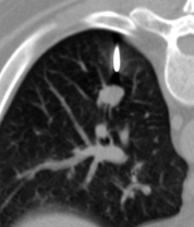
Figure 1. The distance between the lesion and needle.
Before the start of the main stage, the medical staff puts the patient on the table to ensure the best access to the lung. Depending on the location of the formation, it is possible that the patient should be supine resting, prone resting or lie side or lie sidelong. The position is chosen to minimize the pulmonary movement and ensure more accurate insertion of the biopsy needle.
All our studies were conducted mandatory with the local aneasthesia (with or/without sedation). The puncture site was selected individually. Immediately before the start of the procedure, the location of the envisaged intervention is treated with an antiseptic and after the patient was provided with the local anesthetic. During all the time that the patient is in the procedure room, the anesthesiologist monitored the well-being of the patient, as well as pulse rate and blood pressure. The manipulations of the interventional radiologist the insertion and advancement of the needle were controlled via CT.
Once the desired area has been reached, a small sample of lung tissue is removed via a needle. Then, this material was fixed immediately and sent to the pathology laboratory for further microscopic analysis. After successful intake of a tissue sample, the needle is removed for biopsy. The place of the intervention is treated with an antiseptic and covered with a sterile dressing to prevent infection. Before removing the patient from the table a CT scan control to identify possible complications is required. And after this begins the final stage: the patient follow-up.
Despite the fact that we are used to this procedure, and in our department it is carried out quite often, we should not forget about the radiation exposure for the patient and the physician, as well as complications during and after the biopsy.
Even at the stage of preparation for a biopsy, we can assume possible complications. High-risk factors are: females over 65 years old, COPD, smoking, history of vasculitis, lung transplantation, resection of the contralateral lung or presence of pneumonia and flu.
In this regard, we take the following actions: the patient is interviewed in details before the start of the procedure, that includes evaluation of the family and medical history, their habits, alcohol and tobacco use, allergies to any medication, discussion about the risks of anesthesia and the informed consent. Then thorough monitoring of vital signs and saturation of oxygen started, so the patient can be transferred to room tomography. At the end of each procedure the control chest CT scan or chest X-ray are performed. In case of emergency the patient is sent to thoracic surgery department.
The additional time of the observation for the patient is related to the complications to be expected, the severity of the disease and the duration of the procedure. According to the literature, the complications are divided into two categories:
Mild: Observation for 1-4 hours (bleeding, hemoptysis, contusion)
Serious: Emergency, requested the assistance of a thoracic surgeon (bleeding, drainage). The most dangerous are the occurrence of pneumothorax with /without symptoms, hemoptysis (extensive), death.
Table 2. List of complications.

Complications

Number

Pneumothorax (serious)

5 cases

Alveolar hemorrhage (severe)

1 case

Hemoptysis (serious)

3 cases

Death

1 case

Control is performed before 1 hour and during this period constant monitoring of the patient and his/her vital signs by the nurse directly in the department. During the monitoring for 2 to 4 hours, we record all the changes in the breathing, saturation, and pressure. Follow-up 24 hours was performed every 2-4 hours in case of complications identified (local pneumothorax or hemoptisis).
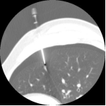
Figure 2. CT-guided transthoracic needle biopsy of a pulmonary lesion.
Pneumothorax: This is a complication that usually occurs immediately after the needle is removed. It is visualized on CTscans in the form of a small strip. Depending on the volume of pneumothorax, we do the following: if the pneumothorax is mild, i.e., less than 15% (the majority is approximately less than 5% - 20%), we perform the control CT and aspiration on the same day just before discharge of the patient. If it exceeds the level (partial or total) the patient is monitored and treated at the thoracic surgery department, and is provided with the intensive monitoring.
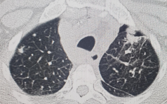
Figure 3. Patient with pneumothorax after the biopsy.
The number of severe complications during the period of 1.5 years was: partial or total pneumothorax - 5.6%, hemorrhage - 1.1%, hemoptysis upto 3.3%.
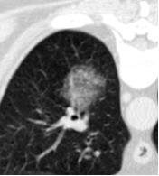
Figure 4. Patient with bleeding after the biopsy.
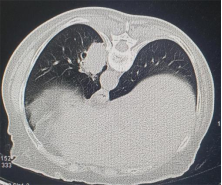
Figure 5. Lesion before the biopsy.
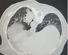
Figure 6. Injury after the biopsy with findings of hemorrhage.
4. Discussion
In our opinion, elevated risk of complications is associated with the long smoking history (clinical data, coagulation disorders). Per the literature data, the percentage of complications that lead to the death of a patient , is about 1%, and in our practice, unfortunately, one case of death was also registered after biopsy. After analysis of this tragedy we came to the conclusion that during the evaluation of each such case, we have to assess not only the risks for the patient, but also we need to use the data from additional diagnostic procedures (e.g., spirometry), when we are able to estimate the extent of response to this type of the disease, and to reduce, as well, the expected and unexpected risks.
5. Conclusion
According to the literature, after a retrospective analysis, the frequency of complications is significantly related to the volume of the lesion, the diameter of the needle, as well as with the depth of the lesion and does not depend on the age or level of training of the physician .
6. Recommendations
Each of these stages of the realization of a lung biopsy guided by CT requires a high degree of professionalism and care from the medical staff. The three stages are important and have the same goal: to make a final diagnosis. Due to the modern methods of and anesthesia, transthoracic biopsy is a relatively safe and minimally invasive procedure that allows to obtain the material required for the diagnosis. The careful planning, the joint work of doctors and nursing staff allows us to minimize the risks, associated with the procedure, and our experience and preparation of the patient for this procedure at all stages reduces the percentage of complications.
Abbreviations

BUN

Blood Urea Nitrogen

CBC

Complete Blood Count

CT

Computer Tomography

HIV

Human Immunodeficiency Virus

MRI

Magnetic Resonance Imaging

PET

Positron Emission Tomography

Author Contributions
Anna Krylova: Conceptualization, Methodology
Kirill Rudinskiy: Writing – original draft, Writing – review & editing
Venecia Alba: Data curation, Resources
Conflicts of Interest
The authors declare absence of conflict of interests.
References
[1] Mir MF, Naik MA, Mali JA. CT guided lung biopsy complications: a single center prospective study. Int J Res Med Sci. 2018 Sep; 6(9): 2946.
[2] Heerink WJ, de Bock GH, de Jonge GJ, et al. Complication rates of CT-guided transthoracic lung biopsy: meta-analysis. Eur Radiol. 2017; 27: 138-148.
[3] Otto S., Mensel B., Friedrich N., et al. Predictors of technical success and rate of complications of image-guided percutaneous transthoracic lung needle biopsy of pulmonary tumors. PLoS One. 2015 Apr 9; 10(4): e0124947.
[4] Dezube R. Transthoracic Needle Biopsy. Johns Hopkins University; Nov 2023.
[5] Villegas J., Moreno L., Piedrahita M. Complications of percutaneous biopsy of lung guided by ct scan. Rev According To Columbus Radiol. 2021; 32(3): 5584-90.
[6] Kharchenko VP, Glagolev NA. Computed tomography X-ray in the diagnosis of diseases of the lungs and the mediastinum. Medika; 2005: 120.
[7] Chernousov AF, Pavlov YV, Pavlov AYu. Diagnosis and treatment of small formations pulmonary peripheral. Surgery. 2010; 6: 61-69.
[8] Ragulin YA, Usachev VS, Medvedev VN, et al. The CT-guided transthoracic biopsy of lung and mediastinal tumors. Medical Radiological Research Center of the Ministry of Health and Social Development of Russia, Obninsk.
[9] Manhire A, Charig M, Clelland C, Gleeson F, Miller R, Moss H, Pointon K, Richardson C, Sawicka E; Guidelines for radiologically guided lung biopsy. Thorax. 2003 Nov; 58 (11): 920-36.
[10] Herf S. M, Suratt P. M, Arora N. S Deaths and complications associated with transbronchial lung biopsy. Am Rev Respir Dis. 1977 Apr; 115(4): 708-11.
[11] Han Y, Kim HJ, Kong KA, Kim SJ, Lee SH, Ryu YJ, Lee JH. Kim Y, Shim SS, Chang JH. Diagnosis of small pulmonary lesions by transbronchial lung biopsy with radial endobronchial ultrasound and virtual bronchoscopic navigation versus CT-guided transthoracic needle biopsy: A systematic review and meta-analysis. PLoS One. 2018 Jan 22; 13(1).
[12] Hu Y, Yang X, Nie L, Zhao D, An J, Li B. Analysis of Clinical Characteristics and Driver Genes in 405 Patients with Lung Cancer Complicated with Tuberculosis. Zhongguo Fei Ai Za Zhi 2020 May 20; 23(5): 337-342.
[13] Jurczak P, Romaszko-Wojtowicz A, Doboszyńska A. Lung Lesions: Differential Diagnoses Beyond Cancer. Am J Case Rep. 2024 Jun 15: 25: e943798.
[14] Ramón Rami-Porta, Hisao Asamura, William D Travis, Valerie W Rusch. Lung cancer - major changes in the American Joint Committee on Cancer eighth edition cancer staging manual CA Cancer J Clin. 2017 Mar; 67(2): 138-155.
[15] Arash Najafi, Marc Al Ahmar, Baptiste Bonnet, Alexandre Delpla, Adrian Kobe, Khaled Madani, Charles Roux, Frédéric Deschamps, Thierry de Baère, Lambros Tselikas. The PEARL Approach for CT-guided Lung Biopsy: Assessment of Complication Rate. Radiology. 2022 Feb; 302(2): 473-480.
Cite This Article
  • APA Style

    Krylova, A., Rudinskiy, K., Alba, V. (2026). A Review of CT-Guided Lung Biopsy: Procedure Stages, Complications, and Risk Mitigation. International Journal of Pharmacy and Chemistry, 12(2), 19-23. https://doi.org/10.11648/j.ijpc.20261202.11

    Copy | Download

    ACS Style

    Krylova, A.; Rudinskiy, K.; Alba, V. A Review of CT-Guided Lung Biopsy: Procedure Stages, Complications, and Risk Mitigation. Int. J. Pharm. Chem. 2026, 12(2), 19-23. doi: 10.11648/j.ijpc.20261202.11

    Copy | Download

    AMA Style

    Krylova A, Rudinskiy K, Alba V. A Review of CT-Guided Lung Biopsy: Procedure Stages, Complications, and Risk Mitigation. Int J Pharm Chem. 2026;12(2):19-23. doi: 10.11648/j.ijpc.20261202.11

    Copy | Download

  • @article{10.11648/j.ijpc.20261202.11,
      author = {Anna Krylova and Kirill Rudinskiy and Venecia Alba},
      title = {A Review of CT-Guided Lung Biopsy: Procedure Stages, Complications, and Risk Mitigation},
      journal = {International Journal of Pharmacy and Chemistry},
      volume = {12},
      number = {2},
      pages = {19-23},
      doi = {10.11648/j.ijpc.20261202.11},
      url = {https://doi.org/10.11648/j.ijpc.20261202.11},
      eprint = {https://article.sciencepublishinggroup.com/pdf/10.11648.j.ijpc.20261202.11},
      abstract = {The modern method of diagnosis of lung lesions, biopsy guided by CT is widely used in clinical practice. Especially this method is used frequently in oncological hospitals. In our study we analyzed data of patients of the oncology institute, who underwent a lung biopsy using a biopsy guided by CTand complications associated with this diagnostic procedure. The aim of our study was to objectively evaluate the types of complications in patients after the procedure, to compare them with data from the preliminary examination in order to reduce or avoid them in the future. Among all patients who underwent this procedure (89), the number of serious complications during this period of 1.5 years was: pneumothorax 5.6%, hemorrhage 1.1%, hemoptisis - upto 3.3%. In our opinion, the elevation of the risks is influenced by long smoking history (clinical data, coagulation disorders). We discuss in detail the tactics of management and observation of patients after the procedure, taking into account the patient's medical history and complications during the procedure. Careful preliminary preparation of the patients before the procedure helps to reduce the risk of complications.},
     year = {2026}
    }
    

    Copy | Download

  • TY  - JOUR
    T1  - A Review of CT-Guided Lung Biopsy: Procedure Stages, Complications, and Risk Mitigation
    AU  - Anna Krylova
    AU  - Kirill Rudinskiy
    AU  - Venecia Alba
    Y1  - 2026/07/03
    PY  - 2026
    N1  - https://doi.org/10.11648/j.ijpc.20261202.11
    DO  - 10.11648/j.ijpc.20261202.11
    T2  - International Journal of Pharmacy and Chemistry
    JF  - International Journal of Pharmacy and Chemistry
    JO  - International Journal of Pharmacy and Chemistry
    SP  - 19
    EP  - 23
    PB  - Science Publishing Group
    SN  - 2575-5749
    UR  - https://doi.org/10.11648/j.ijpc.20261202.11
    AB  - The modern method of diagnosis of lung lesions, biopsy guided by CT is widely used in clinical practice. Especially this method is used frequently in oncological hospitals. In our study we analyzed data of patients of the oncology institute, who underwent a lung biopsy using a biopsy guided by CTand complications associated with this diagnostic procedure. The aim of our study was to objectively evaluate the types of complications in patients after the procedure, to compare them with data from the preliminary examination in order to reduce or avoid them in the future. Among all patients who underwent this procedure (89), the number of serious complications during this period of 1.5 years was: pneumothorax 5.6%, hemorrhage 1.1%, hemoptisis - upto 3.3%. In our opinion, the elevation of the risks is influenced by long smoking history (clinical data, coagulation disorders). We discuss in detail the tactics of management and observation of patients after the procedure, taking into account the patient's medical history and complications during the procedure. Careful preliminary preparation of the patients before the procedure helps to reduce the risk of complications.
    VL  - 12
    IS  - 2
    ER  - 

    Copy | Download

Author Information