Case Report | | Peer-Reviewed

Potential for Corneal Recovery During Nonstandard Rehabilitation After LASIK

Received: 21 July 2026     Accepted: 3 August 2026     Published: 20 August 2026
Views:       Downloads:
Abstract

Laser in situ keratomileusis (LASIK) provides predictable refractive outcomes, but clinically silent epithelial basement membrane dystrophy (EBMD) may become apparent during flap creation and lead to epithelial sloughing, delayed epithelial recovery, and postoperative ocular surface instability. This case report describes the management of a 30-year-old man with bilateral moderate myopia and astigmatism who underwent microkeratome-assisted LASIK after a routine preoperative examination showed no corneal epithelial abnormalities. The baseline Ocular Surface Disease Index score was 6, tear breakup time was 7 seconds in the right eye and 8 seconds in the left eye, and anterior-segment optical coherence tomography showed tear meniscus heights of 304 and 299 micrometers, respectively. During flap repositioning, the epithelium demonstrated abnormal mobility, folding, and focal sloughing. On postoperative day 1, the right eye had uncorrected visual acuity of 0.6-0.7, diffuse epithelial edema, and a central epithelial defect; the left eye had uncorrected visual acuity of approximately 1.0. Subconjunctival dexamethasone was administered because of the risk of diffuse lamellar keratitis. At 1 week, persistent grade 1-2 epitheliopathy and early epithelial ingrowth were observed. Treatment was supplemented with three weekly bilateral subconjunctival injections of 1% low-molecular-weight sodium hyaluronate, followed by monthly injections, together with topical corticosteroids and hyaluronic-acid lubricants. During follow-up, epithelial staining decreased, epithelial morphology on optical coherence tomography became more uniform, visual acuity remained stable, and subjective discomfort improved. At 2 months, the Ocular Surface Disease Index score decreased to 3, Schirmer test values increased to 26 and 28 mm, tear breakup time increased to 9 and 10 seconds, and tear meniscus height increased to 476 and 504 micrometers. This case suggests that multimodal regenerative therapy including subconjunctival low-molecular-weight sodium hyaluronate may support corneal epithelial recovery after intraoperative manifestation of EBMD during LASIK; controlled studies are required to determine efficacy.

Published in Science Journal of Clinical Medicine (Volume 15, Issue 2)
DOI 10.11648/j.sjcm.20261502.14
Page(s) 21-26
Creative Commons

This is an Open Access article, distributed under the terms of the Creative Commons Attribution 4.0 International License (http://creativecommons.org/licenses/by/4.0/), which permits unrestricted use, distribution and reproduction in any medium or format, provided the original work is properly cited.

Copyright

Copyright © The Author(s), 2026. Published by Science Publishing Group

Keywords

LASIK, Epithelial Basement Membrane Dystrophy, Corneal Epithelium, Hyaluronic Acid, Sodium Hyaluronate, Epithelial Sloughing, Ocular Surface, Case Report

1. Introduction
Laser in situ keratomileusis (LASIK) is one of the most frequently performed keratorefractive procedures worldwide . Despite careful patient selection and detailed preoperative assessment, unusual intraoperative findings and atypical postoperative recovery may still occur. Post-refractive ocular surface disturbance, including tear-film instability and dry-eye symptoms, is among the most common early complications and may delay epithelial recovery . Certain corneal disorders, including epithelial basement membrane dystrophy (EBMD), may remain asymptomatic and become clinically apparent only after surgical manipulation.
EBMD is the most common anterior corneal dystrophy, with an estimated prevalence of 2%-6% in the general population . It is characterized by an abnormal basement membrane protruding toward the epithelium and epithelial microcysts . These histologic abnormalities are often clinically silent, which complicates timely diagnosis. In symptomatic cases, recurrent corneal epithelial erosions may develop .
Patients with EBMD have reduced epithelial adhesion and are predisposed to epithelial displacement or sloughing during corneal flap creation . Abnormal hemidesmosome development and deficient anchoring fibrils weaken epithelial attachment and increase the risk of recurrent erosions . Only a limited number of reports describe EBMD becoming manifest during or after keratorefractive surgery .
One regenerative approach used in Belarusian ophthalmic practice is subconjunctival administration of a 1% structured solution of low-molecular-weight sodium hyaluronate (LMW-NaHA; “Gial-In”, HyalSyn Technology, Belarus). Previous clinical and experimental work has reported potential benefit in chronic dystrophic corneal disease and ocular surface dysfunction .
Hyaluronic acid is a glycosaminoglycan composed of repeating D-glucuronic acid and N-acetylglucosamine units. It is present predominantly in the extracellular and pericellular matrices and contributes to tissue hydration, viscoelasticity, cell signaling, cytoskeletal regulation, and the expression of genes involved in proliferation and differentiation . Hyaluronic acid is also involved in proteoglycan supramolecular assembly and in the activation, migration, and differentiation of progenitor cells . The present report describes corneal epithelial recovery after intraoperative manifestation of previously silent EBMD during LASIK and the subsequent use of a multimodal regimen including subconjunctival LMW-NaHA.
2. Case Presentation
2.1. Patient and Preoperative Examination
A 30-year-old man was referred for planned laser vision correction. He reported reduced uncorrected vision and a history of soft contact lens wear. He denied chronic systemic disease, recurrent herpetic infection, and continuous medication use. His occupational history included approximately 8 hours of computer work daily. A comprehensive ophthalmic and systemic examination was performed to exclude contraindications to refractive surgery.
Best-corrected distance visual acuity was determined using an optometric protocol that accounted for refractive and accommodative-muscular balance. In the right eye, uncorrected distance visual acuity (UDVA) was 0.1 and best-corrected distance visual acuity (BCDVA) was 1.0 with -5.50 D sphere and -1.00 D cylinder at 170 degrees. In the left eye, UDVA was 0.1 and BCDVA was 1.0 with -5.50 D sphere and -0.75 D cylinder at 175 degrees. The spherical equivalents were -6.00 D and -6.12 D, respectively. Additional preoperative findings are summarized in Table 1.
The patient reported occasional foreign-body sensation, photophobia, and discomfort in windy conditions. His Ocular Surface Disease Index (OSDI) score was 6, indicating minimal symptoms of disturbed ocular surface homeostasis. Slit-lamp examination showed pale-pink conjunctiva and clear, spherical, lustrous corneas. Fluorescein staining did not reveal epithelial abnormalities. Examination of the remaining ocular media and retina disclosed no contraindication to laser correction. The clinical diagnosis was bilateral moderate myopia with astigmatism and bilateral chorioretinal pigmentary degeneration.
Table 1. Preoperative clinical characteristics.

Parameter

OD

OS

UDVA/BCDVA

0.1/1.0

0.1/1.0

Autokeratorefractometry, D

-5.50 -1.00 x 170°

-5.50 -0.75 x 175°

Spherical equivalent, D

-6.00

-6.12

Keratometry K1/K2, D @ axis

43.46 @ 173° / 44.47 @ 83°

43.31 @ 176° / 44.27 @ 86°

Schirmer test, mm

20

15

Central pachymetry, µm

540

543

TBUT, s

7

8

Meibomian gland loss, upper/lower lid,%

5/13.7 (grade 1)

11.4/22.9 (grade 1)

Tear meniscus height by AS-OCT, µm

304

299

OD, right eye; OS, left eye; UDVA, uncorrected distance visual acuity; BCDVA, best-corrected distance visual acuity; TBUT, tear breakup time; AS-OCT, anterior-segment optical coherence tomography.
2.2. LASIK Procedure and Early Postoperative Course
LASIK was performed in both eyes. A 90-µm corneal flap was created with a Moria 3One Use-Plus microkeratome (Moria, France). Stromal ablation was performed with a SCHWIND AMARIS 500E excimer laser (SCHWIND eye-tech-solutions, Germany). The planned ablation-zone diameters were approximately 8.19 mm in the right eye and 8.15 mm in the left eye, taking pupillometry into account. Ablation depth was 103 µm in the right eye and 98 µm in the left eye.
Flap creation and excimer-laser ablation were completed without a mechanical flap complication. However, during flap repositioning, the epithelium demonstrated abnormal mobility relative to Bowman membrane, formed folds, and detached in small fragments. Postoperatively, topical antimicrobial therapy and lubricants were prescribed.
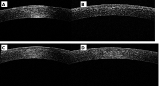
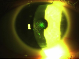
On postoperative day 1, UDVA was 0.6-0.7 in the right eye and approximately 1.0 in the left eye. The patient reported blurred vision, discomfort, and photophobia. Slit-lamp examination showed diffuse epithelial edema and a central fluorescein-positive epithelial defect in the right eye (Figure 1). Anterior-segment OCT showed marked epithelial irregularity in both eyes (Figure 5A and 5B).
Figure 1. Fluorescein-stained anterior segment of the right eye on postoperative day 1 after LASIK, showing a central epithelial defect.
Because of pronounced edema of the anterior corneal layers and the perceived risk of diffuse lamellar keratitis, 0.3 mL of dexamethasone was administered subconjunctivally. Topical anti-inflammatory therapy was continued according to the postoperative protocol.
2.3. Regenerative Treatment and Follow-up
At the 1-week visit, blurred vision, discomfort, and photophobia persisted. Visual acuity was 1.0 in the right eye and 1.2 in the left eye. Manifest refraction was +0.25 D sphere with +0.75 D cylinder at 46 degrees in the right eye and +0.50 D sphere with -0.75 D cylinder at 126 degrees in the left eye; the corresponding spherical equivalents were +0.63 D and +0.13 D. Epithelial edema had decreased, but fluorescein staining showed grade 1-2 epitheliopathy according to the Efron grading scale. Persistent epitheliopathy and early epithelial ingrowth at the inferior flap edge were interpreted as signs of impaired ocular surface homeostasis and active inflammation associated with EBMD manifestation.
The regimen was supplemented with 1% LMW-NaHA (structured solution), administered subconjunctivally at 0.5 mL in each eye once weekly for three injections. Topical corticosteroids and lubricants were continued . The patient remained under close observation.
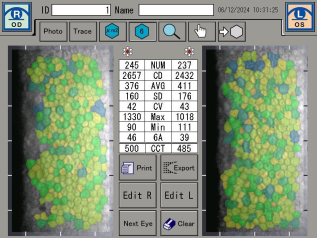
Figure 2. Specular endothelial microscopy of both eyes. No clinically significant endothelial abnormalities were identified.
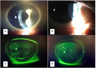
At 1 month, the right eye showed minimal epithelial ingrowth at the inferior flap edge without signs of continued progression and only grade 0-1 epitheliopathy. In the left eye, the flap was well adapted; at the 6-o’clock position, minimal fluorescein staining corresponded to grade 1 peripheral epitheliopathy (Figure 3).
Specular endothelial microscopy was performed to further evaluate the corneal endothelium (Figure 2). No pathologic endothelial changes were detected, allowing attention to be focused on regenerative processes in the anterior corneal layers, including the epithelium, epithelial basement membrane, Bowman membrane, and superficial stroma. Continued instillation of 0.4% hyaluronic-acid lubricant and monthly bilateral subconjunctival injections of 1% LMW-NaHA at 0.3 mL per eye were recommended.
Figure 3. Anterior segment 1 month after LASIK. The right eye (A, C) shows more pronounced fluorescein staining, consistent with weaker epithelial adhesion to the basement membrane. The left eye (B, D) shows less staining and a more favorable healing pattern.
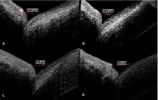
Figure 4. Tear meniscus height before surgery (A, B) and 2 months after surgery (C, D). Measurements increased from 304 and 299 µm to 476 and 504 µm in the right and left eyes, respectively.
Subsequent examinations demonstrated stable visual acuity and resolution of the patient’s discomfort. Mild grade 1-2 inferior corneal epitheliopathy with limited fluorescein staining remained, but no signs of epithelial adhesion failure were observed. Anterior-segment OCT showed a more uniform epithelial profile (Figure 5). The OSDI score decreased to 3; the patient reported only rare discomfort after sleep.
Objective ocular surface parameters also improved. Schirmer test values increased to 26 mm in the right eye and 28 mm in the left eye; TBUT increased to 9 and 10 seconds; and tear meniscus height on OCT increased relative to baseline (Figure 4). Continued monthly regenerative therapy and topical hyaluronic-acid instillation were recommended to maintain the result and reduce factors that could provoke recurrent epithelial detachment.
Figure 5. Anterior-segment OCT of both corneas on postoperative day 1 (A, B) and at 2 months (C, D), demonstrating improved epithelial regularity during follow-up.
3. Discussion
This case is clinically relevant because silent EBMD may remain undetected despite comprehensive routine preoperative assessment and may first become apparent during refractive surgery. Standard slit-lamp examination and fluorescein staining were normal before surgery, yet flap repositioning immediately revealed abnormal epithelial mobility and focal sloughing. In vivo confocal microscopy can help identify subtle abnormalities of the epithelial basement membrane and subbasal nerve plexus, but it is not universally available. Recent optical approaches based on corneal epithelial wavefront analysis may improve detection of subtle EBMD-related irregularity that is not evident on routine examination Consequently, careful intraoperative observation and postoperative multimodal imaging remain important when recovery is atypical.
The patient was monitored with slit-lamp biomicroscopy, fluorescein staining, AS-OCT, specular endothelial microscopy, Schirmer testing, TBUT, tear meniscus measurements, and the OSDI questionnaire. Contemporary reviews of post-refractive ocular surface disease emphasize baseline and serial assessment of tear-film stability, epithelial integrity, and patient-reported symptoms . This combination allowed simultaneous assessment of epithelial integrity, corneal morphology, tear-film stability, and subjective symptoms. The absence of endothelial abnormalities helped localize the clinically relevant process to the anterior corneal layers.
The therapeutic approach combined topical corticosteroids and lubricants with subconjunctival LMW-NaHA. Hyaluronic acid may contribute to tissue hydration, extracellular-matrix organization, cell migration, and epithelial repair . In the present patient, treatment was temporally associated with reduced fluorescein staining, improved epithelial regularity on OCT, stable BCDVA without loss of lines, lower OSDI score, increased Schirmer test values, longer TBUT, and increased tear meniscus height. These findings are consistent with previous reports describing improvement in ocular surface homeostasis with hyaluronic-acid-based therapy .
Published cases of EBMD manifesting after LASIK have sometimes required photorefractive or phototherapeutic keratectomy to treat persistent epithelial irregularity or recurrent symptoms . The present case was managed conservatively and did not require surface ablation during the reported observation period. Nevertheless, a single uncontrolled case cannot establish causality or comparative effectiveness. Spontaneous epithelial recovery, topical corticosteroid therapy, lubrication, and close follow-up may also have contributed to the favorable course. Prospective controlled studies are needed to determine the independent efficacy, optimal dose, safety, and duration of subconjunctival LMW-NaHA in postoperative epithelial disorders.
4. Conclusions
Previously silent EBMD may present intraoperatively during LASIK as excessive epithelial mobility, folding, and focal sloughing despite an unremarkable routine preoperative examination. In this patient, a multimodal conservative regimen that included topical corticosteroids, hyaluronic-acid lubricants, and subconjunctival 1%structured LMW-NaHA was associated with progressive epithelial stabilization, improved ocular surface parameters, preserved visual acuity, and reduced symptoms. This approach may offer a nonsurgical option during early rehabilitation, but its efficacy and safety require confirmation in larger controlled studies.
5. Recommendations
Epithelial basement membrane dystrophy remains a clinically relevant problem in refractive surgery because mild or asymptomatic forms may not be detected during routine preoperative examination. Standard slit-lamp biomicroscopy and fluorescein staining may be insufficient to identify subtle abnormalities of epithelial adhesion, while more sensitive diagnostic methods, including in vivo confocal microscopy, are not universally available in routine clinical practice.
Further research should therefore focus on improving preoperative identification of patients at risk of epithelial instability and on developing accessible methods for monitoring corneal epithelial and basement membrane recovery after refractive surgery. Particular attention should be given to multimodal assessment combining clinical examination, fluorescein staining, anterior-segment optical coherence tomography, tear-film parameters, and patient-reported symptoms.
An additional priority is the investigation of regenerative treatment strategies that can support corneal healing with minimal cytotoxic effects. Prospective controlled studies are required to evaluate the efficacy, safety, optimal dosage, administration schedule, and long-term outcomes of subconjunctival structured low-molecular-weight sodium hyaluronate and other regenerative agents. Our research group is currently developing and refining clinical algorithms for the use of these regenerative approaches in order to achieve the best possible anatomical, functional, and patient-reported outcomes. Such studies may help make postoperative corneal recovery more predictable and controllable, including in patients with atypical or complicated healing, while preserving visual function and reducing the need for additional surgical intervention.
Abbreviations

AS-OCT

Anterior-segment Optical Coherence Tomography

BCDVA

Best-corrected Distance Visual Acuity

EBMD

Epithelial Basement Membrane Dystrophy

LASIK

Laser in Situ Keratomileusis

LMW-NaHA

Low-molecular-weight Sodium Hyaluronate

OD

Right Eye

OS

Left Eye

OSDI

Ocular Surface Disease Index

TBUT

Tear Breakup Time

UDVA

Uncorrected Distance Visual Acuity

Author Contributions
Galina Romanovna Semak: Conceptualization, Supervision, Writing – review & editing
Volha Ivanovna Remezava: Data curation, Formal Analysis, Investigation, Writing – original draft
Yuliya Vladimirovna Markava: Investigation, Validation, Visualization, Writing – original draft
Gleb Valerievich Krishtopenko: Writing – review & editing
Data Availability Statement
The data supporting the outcome of this case report are presented in the manuscript.
Conflicts of Interest
The authors declare no conflicts of interest.
References
[1] Sahay P, Bafna RK, Reddy JC, Vajpayee RB, Sharma N. Complications of laser-assisted in situ keratomileusis. Indian Journal of Ophthalmology. 2021; 69(7): 1658-1669.
[2] Laibson PR. Microcystic corneal dystrophy. Trans Am Ophthalmol Soc. 1976; 74: 488–531.
[3] Labbé A, Nicola RD, Dupas B, Auclin F, Baudouin C. Epithelial basement membrane dystrophy: evaluation with the HRT II Rostock Cornea Module. Ophthalmology. 2006 Aug; 113(8): 1301-8.
[4] Semak GR. Activation of Regeneration and Hyaluronic Acid in Corneal Dystrophy. Minsk: Belarusian State Medical University; 2023. 171 p.
[5] Dastgheib A, Thomas E, Manche E. Sloughing of corneal epithelium and wound healing complications associated with laser in situ keratomileusis in patients with epithelial basement membrane dystrophy. American Journal of Ophthalmology. 2000; 130(3): 297-303.
[6] Pérez-Santonja JJ, Galal A, Cardona C, Artola A, Ruíz-Moreno JM, Alió JL. Severe corneal epithelial sloughing during laser in situ keratomileusis as a presenting sign for silent epithelial basement membrane dystrophy. Journal of Cataract & Refractive Surgery. 2005; 31(10): 1932-1937.
[7] Surnina ZV, Sukhanova EV, Gamidov AA. Clinical manifestation of corneal epithelial dystrophy after LASIK. Medical Council. 2022; 16(23): 275-283. (In Russian).
[8] Wu PY, Tsui MC, Chang CK, Chang HW, Chen WL. Epithelial basement membrane dystrophy after femtosecond laser-assisted LASIK successfully treated with in vivo confocal microscopy-assisted photorefractive keratectomy. Journal of Cataract & Refractive Surgery. 2020; 46(12): e17-e19.
[9] Semak GR. Clinical and Experimental Substantiation of Pathogenetic Methods for Treating Chronic Dystrophic Corneal Diseases [doctoral dissertation]. Minsk; 2022. 222 p. (In Russian).
[10] Semak G, Ryndova D, Smiantsina A, Kastsiukovich Y. Restoration of ocular surface homeostasis after excimer laser correction. Ophthalmology. Eastern Europe. 2023; 13(2): 97-109.
[11] Zhu J, Tang X, Ho CT, Huang Q. Applications and delivery mechanisms of hyaluronic acid used for topical/transdermal delivery. International Journal of Pharmaceutics. 2020; 578: 119127.
[12] Woreta FA, Davis GW, Bower KS. LASIK and surface ablation in corneal dystrophies. Survey of Ophthalmology. 2015; 60(2): 115-122.
[13] Nair S, Kaur M, Sharma N, Titiyal JS. Refractive surgery and dry eye - An update. Indian Journal of Ophthalmology. 2023; 71(4): 1105-1114.
[14] Kim BK, Chung YT. Clinical outcomes of epithelial basement membrane dystrophy after keratorefractive lenticule extraction. Journal of Cataract & Refractive Surgery. 2024; 50(6): 558-564.
[15] Grauvogl V, Mayer WJ, Siedlecki J, Mohr N, Dirisamer M, Priglinger SG, Kassumeh S, Luft N. Corneal Epithelial Wavefront Error as a Novel Diagnostic Marker for Epithelial Basement Membrane Dystrophy. Life. 2024; 14(9): 1188.
Cite This Article
  • APA Style

    Semak, G. R., Remezava, V. I., Markava, Y. V., Krishtopenko, G. V. (2026). Potential for Corneal Recovery During Nonstandard Rehabilitation After LASIK. Science Journal of Clinical Medicine, 15(2), 21-26. https://doi.org/10.11648/j.sjcm.20261502.14

    Copy | Download

    ACS Style

    Semak, G. R.; Remezava, V. I.; Markava, Y. V.; Krishtopenko, G. V. Potential for Corneal Recovery During Nonstandard Rehabilitation After LASIK. Sci. J. Clin. Med. 2026, 15(2), 21-26. doi: 10.11648/j.sjcm.20261502.14

    Copy | Download

    AMA Style

    Semak GR, Remezava VI, Markava YV, Krishtopenko GV. Potential for Corneal Recovery During Nonstandard Rehabilitation After LASIK. Sci J Clin Med. 2026;15(2):21-26. doi: 10.11648/j.sjcm.20261502.14

    Copy | Download

  • @article{10.11648/j.sjcm.20261502.14,
      author = {Galina Romanovna Semak and Volha Ivanovna Remezava and Yuliya Vladimirovna Markava and Gleb Valerievich Krishtopenko},
      title = {Potential for Corneal Recovery During Nonstandard Rehabilitation After LASIK},
      journal = {Science Journal of Clinical Medicine},
      volume = {15},
      number = {2},
      pages = {21-26},
      doi = {10.11648/j.sjcm.20261502.14},
      url = {https://doi.org/10.11648/j.sjcm.20261502.14},
      eprint = {https://article.sciencepublishinggroup.com/pdf/10.11648.j.sjcm.20261502.14},
      abstract = {Laser in situ keratomileusis (LASIK) provides predictable refractive outcomes, but clinically silent epithelial basement membrane dystrophy (EBMD) may become apparent during flap creation and lead to epithelial sloughing, delayed epithelial recovery, and postoperative ocular surface instability. This case report describes the management of a 30-year-old man with bilateral moderate myopia and astigmatism who underwent microkeratome-assisted LASIK after a routine preoperative examination showed no corneal epithelial abnormalities. The baseline Ocular Surface Disease Index score was 6, tear breakup time was 7 seconds in the right eye and 8 seconds in the left eye, and anterior-segment optical coherence tomography showed tear meniscus heights of 304 and 299 micrometers, respectively. During flap repositioning, the epithelium demonstrated abnormal mobility, folding, and focal sloughing. On postoperative day 1, the right eye had uncorrected visual acuity of 0.6-0.7, diffuse epithelial edema, and a central epithelial defect; the left eye had uncorrected visual acuity of approximately 1.0. Subconjunctival dexamethasone was administered because of the risk of diffuse lamellar keratitis. At 1 week, persistent grade 1-2 epitheliopathy and early epithelial ingrowth were observed. Treatment was supplemented with three weekly bilateral subconjunctival injections of 1% low-molecular-weight sodium hyaluronate, followed by monthly injections, together with topical corticosteroids and hyaluronic-acid lubricants. During follow-up, epithelial staining decreased, epithelial morphology on optical coherence tomography became more uniform, visual acuity remained stable, and subjective discomfort improved. At 2 months, the Ocular Surface Disease Index score decreased to 3, Schirmer test values increased to 26 and 28 mm, tear breakup time increased to 9 and 10 seconds, and tear meniscus height increased to 476 and 504 micrometers. This case suggests that multimodal regenerative therapy including subconjunctival low-molecular-weight sodium hyaluronate may support corneal epithelial recovery after intraoperative manifestation of EBMD during LASIK; controlled studies are required to determine efficacy.},
     year = {2026}
    }
    

    Copy | Download

  • TY  - JOUR
    T1  - Potential for Corneal Recovery During Nonstandard Rehabilitation After LASIK
    AU  - Galina Romanovna Semak
    AU  - Volha Ivanovna Remezava
    AU  - Yuliya Vladimirovna Markava
    AU  - Gleb Valerievich Krishtopenko
    Y1  - 2026/08/20
    PY  - 2026
    N1  - https://doi.org/10.11648/j.sjcm.20261502.14
    DO  - 10.11648/j.sjcm.20261502.14
    T2  - Science Journal of Clinical Medicine
    JF  - Science Journal of Clinical Medicine
    JO  - Science Journal of Clinical Medicine
    SP  - 21
    EP  - 26
    PB  - Science Publishing Group
    SN  - 2327-2732
    UR  - https://doi.org/10.11648/j.sjcm.20261502.14
    AB  - Laser in situ keratomileusis (LASIK) provides predictable refractive outcomes, but clinically silent epithelial basement membrane dystrophy (EBMD) may become apparent during flap creation and lead to epithelial sloughing, delayed epithelial recovery, and postoperative ocular surface instability. This case report describes the management of a 30-year-old man with bilateral moderate myopia and astigmatism who underwent microkeratome-assisted LASIK after a routine preoperative examination showed no corneal epithelial abnormalities. The baseline Ocular Surface Disease Index score was 6, tear breakup time was 7 seconds in the right eye and 8 seconds in the left eye, and anterior-segment optical coherence tomography showed tear meniscus heights of 304 and 299 micrometers, respectively. During flap repositioning, the epithelium demonstrated abnormal mobility, folding, and focal sloughing. On postoperative day 1, the right eye had uncorrected visual acuity of 0.6-0.7, diffuse epithelial edema, and a central epithelial defect; the left eye had uncorrected visual acuity of approximately 1.0. Subconjunctival dexamethasone was administered because of the risk of diffuse lamellar keratitis. At 1 week, persistent grade 1-2 epitheliopathy and early epithelial ingrowth were observed. Treatment was supplemented with three weekly bilateral subconjunctival injections of 1% low-molecular-weight sodium hyaluronate, followed by monthly injections, together with topical corticosteroids and hyaluronic-acid lubricants. During follow-up, epithelial staining decreased, epithelial morphology on optical coherence tomography became more uniform, visual acuity remained stable, and subjective discomfort improved. At 2 months, the Ocular Surface Disease Index score decreased to 3, Schirmer test values increased to 26 and 28 mm, tear breakup time increased to 9 and 10 seconds, and tear meniscus height increased to 476 and 504 micrometers. This case suggests that multimodal regenerative therapy including subconjunctival low-molecular-weight sodium hyaluronate may support corneal epithelial recovery after intraoperative manifestation of EBMD during LASIK; controlled studies are required to determine efficacy.
    VL  - 15
    IS  - 2
    ER  - 

    Copy | Download

Author Information
  • Department of Ophthalmology, Belarusian State Medical University, Minsk, Belarus

  • Department of Ophthalmology, Branch of Open Joint Stock Company Minsk Tractor Works - Medical Center MTZ, Minsk, Belarus
  • Department of Ophthalmology, Branch of Open Joint Stock Company Minsk Tractor Works - Medical Center MTZ, Minsk, Belarus

  • Department of Ophthalmology, Branch of Open Joint Stock Company Minsk Tractor Works - Medical Center MTZ, Minsk, Belarus