
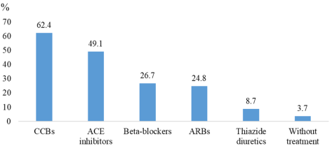
Introduction and objective: Hypertension is a major global public health problem and is a leading risk factor for cardiovascular diseases worldwide due to its prevalence and complications. The objective of this study was to determine the electrocardiographic and echocardiographic characteristics of hypertensive patients at the Renaissance University Hospital Center in N'Djamena, Chad. Patients and Methods: This was a descriptive cross-sectional study with retrospective data collection over a 12-month period from June 1st, 2023 to May 31st, 2024. The study included all medical records of hypertensive patients aged 18 years and older who were on follow up in the Cardiology Department and that underwent an electrocardiogram and a Doppler echocardiogram. We examined sociodemographic, clinical, paraclinical, and therapeutic variables. Results: A total of 322 records were collected. The patient population was predominantly male, with a male-to-female sex ratio of 1.3. The mean age was 55.6 years. Civil servants accounted for 33.2% of cases. The main cardiovascular risk factors identified were diabetes (21.4%), obesity (21%), and dyslipidemia (18.6%). Functionally, Dieulafoy’s neurosensory signs were the most common, at 52.4%. The most commonly prescribed antihypertensive drugs were calcium channel blockers (62.4%) and angiotensin-converting enzyme inhibitors (49.1%). On electrocardiogram, the most common abnormalities were atrial hypertrophy (16.1%) and ventricular hypertrophy (13.6%). Cardiac arrhythmia due to atrial fibrillation was observed in 3.4% of cases. On echocardiography, the most common abnormalities were ventricular hypertrophy (32.3%), left atrial dilation (14.6%), and reduced left ventricular systolic function (7.1%). Conclusion: In this study, electrocardiographic and echocardiographic abnormalities in hypertensive patients were dominated by left ventricular hypertrophy, left atrial dilation, and left ventricular diastolic dysfunction. These abnormalities were most common in patients with long-standing, uncontrolled hypertension.
| Published in | Cardiology and Cardiovascular Research (Volume 10, Issue 3) |
| DOI | 10.11648/j.ccr.20261003.14 |
| Page(s) | 49-54 |
| Creative Commons |
This is an Open Access article, distributed under the terms of the Creative Commons Attribution 4.0 International License (http://creativecommons.org/licenses/by/4.0/), which permits unrestricted use, distribution and reproduction in any medium or format, provided the original work is properly cited. |
| Copyright |
Copyright © The Author(s), 2026. Published by Science Publishing Group |
Electrocardiogram, Echocardiography, Hypertension, N’Djamena
Settings | Number (n) | Percentage (%) |
|---|---|---|
Sex | ||
Men | 137 | 43 |
Women | 185 | 57 |
Age (year) | ||
Means | 55.6 | |
18 – 29 | 3 | 0.9 |
30 – 39 | 41 | 12.7 |
40 – 49 | 69 | 21.4 |
50 – 59 | 99 | 30.7 |
60 et plus | 110 | 32.4 |
Profession | ||
Civil servant | 107 | 33.2 |
Commerçant | 39 | 12.1 |
Farmer | 2 | 0.6 |
Housewife | 97 | 30.1 |
Other | 77 | 23.9 |
Cardiovascular risk factors | ||
Diabetes | 69 | 21.4 |
Obesity | 67 | 21 |
Dyslipidemia | 60 | 18.6 |
Smoking | 16 | 5 |
Kidney failure | 9 | 3 |
Settings | Number (n) | Percentage (%) |
|---|---|---|
Duration (years) | ||
< 1 | 107 | 33.2 |
1 – 2 | 35 | 10.9 |
3 – 4 | 68 | 21.1 |
≥ 5 | 112 | 34.8 |
Severity | ||
Controlled | 90 | 28 |
Grade 1 | 99 | 30.7 |
Grade 2 | 71 | 22 |
Grade 3 | 62 | 19.3 |
Patient follow up | ||
Yes | 191 | 59.3 |
No | 131 | 40.7 |
Therapeutic adherence | ||
Regular | 136 | 42.2 |
Irregular | 186 | 57.8 |
Settings | Number (n) | Percentage (%) |
|---|---|---|
Arrhythmia | ||
Atrial fibrillation | 11 | 3.4 |
Atrial extrasystole | 6 | 2 |
Atrial flutter | 2 | 0.6 |
Ventricular extrasystole | 15 | 4.6 |
Conduction disorder | ||
1st degree AV block | 21 | 6.5 |
2nd degree AV block | 3 | 0.9 |
3rd degree AV block | 2 | 0.6 |
Hypertrophy | ||
Left atrial appendage | 52 | 16.1 |
Right atrial appendage | 3 | 0.9 |
Left ventricular | 44 | 13.6 |
Right ventricular | 1 | 0.3 |
Settings | Number (n) | Percentage (%) |
|---|---|---|
LVEF (%) | ||
<40 | 23 | 7.1 |
40–49 | 17 | 5.3 |
>50 | 282 | 87.6 |
LV Diastolic Dysfunction | ||
Yes | 188 | 58.4 |
No | 134 | 41.6 |
Cavity dilation | ||
Left atrium | 47 | 14.6 |
Right atrium | 9 | 2.7 |
Left ventricle | 33 | 10.2 |
Right ventricle | 4 | 1.2 |
Proximal aorta | 4 | 1.2 |
LV hypertrophy | ||
Concentric | 55 | 17.1 |
Eccentric | 49 | 15.2 |
LVH | Duration of hypertension (years) | p | ||||
|---|---|---|---|---|---|---|
< 1 | 1 - 2 | 3 - 4 | ≥ 5 | Total | ||
Yes | 33 | 5 | 21 | 45 | 104 | 0.03 |
No | 74 | 30 | 47 | 67 | 218 | |
Total | 107 | 35 | 68 | 112 | 322 | |
ECG | Electrocardiogram |
BNP | Brain Natriuretic Peptide |
CCBs | Calcium Channel Blockers |
ACE | Angiotensin-Converting Enzyme |
ARBs | Angiotensin II Receptor Blockers |
LV | Left Ventricular |
LVH | Left Ventricular Hypertrophy |
EF | Ejection Fraction |
| [1] | Ouédraogo S, Zan AA, Ouédraogo EWM, et al. Profile of Hypertension in a Regional Hospital in Burkina Faso. Health 2021; Sci. Dis: 22(12): 116-22. |
| [2] | Sarr SA, Babaka K, Mboup MC, et al. Clinical, electrocardiographic, and echocardiographic aspects of hypertension in the elderly in Senegal. Pan Afr Med J. 2016; 25: 77. |
| [3] | Hamoudi A, Abdelaziz AB, Melki S, et al. Epidemiology of hypertension in Tunisia. The Hammam Sousse/Sahloul Heart Study (HSHS). Tunis Med. 2022 100(2): 167–79. |
| [4] | Michel B, Hermann Y, Liliane KJ, et al. A Study of Left Ventricular Geometry in Black Africans with Hypertension. Annals of Cardiology and Angiology 2023; 72(2): 101–583. |
| [5] | Ali AA, Kaboré A, Doune N, et al. Cross-sectional study on the epidemiological characteristics of hypertension in the context of mass screening conducted among adult volunteers at the Renaissance University Hospital Center. 2022; 6(1): 1-4. |
| [6] | Abdelmadjid Z, Ahamat A, Fouksouna S, et al. Prevalence Hospital of Arterial Hypertension in the Cardiology Departement of the National Reference of Teaching Hopspital. CCR. 2023; 5(2): 80-94. |
| [7] | Ali AA, Doune N, Kaboré A, et al. Prevalence of hypertension in N'Djamena, Chad: Screening campaign among adult volunteers from the general population J Cardio Res. 2023; 188(25): 3-4. |
| [8] | Lang RM, Badano LP, Mor-Avi V, et al. Recommendations for cardiac chamber quantification by echocardiography in adults: an update from the American Society of Echocardiography and the European Association of Cardiovascular Imaging. J Am Soc Echocardiogr 2015; 28: 1-39. |
| [9] | Abdelmadjid ZZ, Ahmat AA, Sakadi F, et al. Prevalence Hospital of arterial hypertension in the Cardiology Department of the National Reference of Teaching Hospital. Ccr 2021; 5(2): 80. |
| [10] | Brouri M, Ouadahi N, Nibouche D, and al. Cardiovascular risk factors in Algeria. A subgroup analysis of the ARICA/Middle East Cardiovascular Epidemiological Study. Annals of Cardiology and Angiology 2018; 67(2): 61-66. |
| [11] | Benyaich A, Aksissou M, Hormi A, et al. Assessment of risk factors for chronic noncommunicable diseases in the rural area of Oujda-Angad, Morocco. Medicine of Metabolic Diseases, 2025; 19(2): 179-191. |
| [12] | Jacques KK, Alain KWM, Grace KN, et al. Prevalence of Hypertension in a Hospital Workplace: The Case of the Kamina General Referral Hospital in the DRC 2023; 25(7): 74–78. |
| [13] | El Boukhrissi F, Bamou Y, Ouleghzal H, et al. Prevalence of risk factors for cardiovascular disease and metabolic syndrome among women in the Meknes region, Morocco. Medicine of Metabolic Diseases 2017; 11(2): 188-194. |
| [14] | Sako M, Konaté M, Sidibé S, et al. Hypertension in the Cardiology Department of Point G University Hospital (Bamako). Health Sci. Dis.: 2024; 25(4): 46–48. |
| [15] | Levy D, Labib SB, Anderson KM, et al. Determinants of sensitivity and specificity of electrocardiographiccriteria for left ventricular hypertrophy. Circulation 1990; 81: 815-82. |
| [16] | Pose-Reino A, González J JR, Pastor C, et al. Myocardial structure and study of arrhythmias during ambulatory electrocardiographic monitoring in mild essential hypertension. Med Clin (Barc). 1996 Jan; 106(1): 7-10. |
| [17] | Nicolas B. Dayal and Hajo Muller. Detection of left ventricular hypertrophy on echocardiography in adults. Rev Med Suisse 2017; 13: 1106-12. |
| [18] | Cissoko AS, Touré M, Dembelé B, et al. Hypertension in the Elderly at Gabriel Touré University Hospital (Bamako). Health Sci. Dis. 2022; 23(8): 112–115. |
| [19] | Mbaye A, Dodo B, Ngaïde AA, et al. Left ventricular hypertrophy in hypertensive Black Africans: results of a cross-sectional study conducted in a semi-rural area of Senegal. 2017; 66(4): 210-16. |
| [20] | Verdecchia P, Schillaci G, Borgioni C, et al. Prognostic significance of serial changes in left ventricular mass in essential hypertension Circulation 1998; 97: 48-54. |
APA Style
Ahamat, A. A., Narcisse, D., Adelroudjal, M. S., Aware, B., Urbain, H., et al. (2026). Electrocardiographic and Echocardiographic Aspects of Hypertensive Patients at the Renaissance University Hospital Center in N'Djamena, Chad. Cardiology and Cardiovascular Research, 10(3), 49-54. https://doi.org/10.11648/j.ccr.20261003.14
ACS Style
Ahamat, A. A.; Narcisse, D.; Adelroudjal, M. S.; Aware, B.; Urbain, H., et al. Electrocardiographic and Echocardiographic Aspects of Hypertensive Patients at the Renaissance University Hospital Center in N'Djamena, Chad. Cardiol. Cardiovasc. Res. 2026, 10(3), 49-54. doi: 10.11648/j.ccr.20261003.14
AMA Style
Ahamat AA, Narcisse D, Adelroudjal MS, Aware B, Urbain H, et al. Electrocardiographic and Echocardiographic Aspects of Hypertensive Patients at the Renaissance University Hospital Center in N'Djamena, Chad. Cardiol Cardiovasc Res. 2026;10(3):49-54. doi: 10.11648/j.ccr.20261003.14
@article{10.11648/j.ccr.20261003.14,
  author = {Ali Adam Ahamat and Doune Narcisse and Mariam Seid Adelroudjal and Brahim Aware and Houba Urbain and Mbaidedjim Sylvain and Yusra Aboulbachar},
  title = {Electrocardiographic and Echocardiographic Aspects of Hypertensive Patients at the Renaissance University Hospital Center in N'Djamena, Chad},
  journal = {Cardiology and Cardiovascular Research},
  volume = {10},
  number = {3},
  pages = {49-54},
  doi = {10.11648/j.ccr.20261003.14},
  url = {https://doi.org/10.11648/j.ccr.20261003.14},
  eprint = {https://article.sciencepublishinggroup.com/pdf/10.11648.j.ccr.20261003.14},
  abstract = {Introduction and objective: Hypertension is a major global public health problem and is a leading risk factor for cardiovascular diseases worldwide due to its prevalence and complications. The objective of this study was to determine the electrocardiographic and echocardiographic characteristics of hypertensive patients at the Renaissance University Hospital Center in N'Djamena, Chad. Patients and Methods: This was a descriptive cross-sectional study with retrospective data collection over a 12-month period from June 1st, 2023 to May 31st, 2024. The study included all medical records of hypertensive patients aged 18 years and older who were on follow up in the Cardiology Department and that underwent an electrocardiogram and a Doppler echocardiogram. We examined sociodemographic, clinical, paraclinical, and therapeutic variables. Results: A total of 322 records were collected. The patient population was predominantly male, with a male-to-female sex ratio of 1.3. The mean age was 55.6 years. Civil servants accounted for 33.2% of cases. The main cardiovascular risk factors identified were diabetes (21.4%), obesity (21%), and dyslipidemia (18.6%). Functionally, Dieulafoy’s neurosensory signs were the most common, at 52.4%. The most commonly prescribed antihypertensive drugs were calcium channel blockers (62.4%) and angiotensin-converting enzyme inhibitors (49.1%). On electrocardiogram, the most common abnormalities were atrial hypertrophy (16.1%) and ventricular hypertrophy (13.6%). Cardiac arrhythmia due to atrial fibrillation was observed in 3.4% of cases. On echocardiography, the most common abnormalities were ventricular hypertrophy (32.3%), left atrial dilation (14.6%), and reduced left ventricular systolic function (7.1%). Conclusion: In this study, electrocardiographic and echocardiographic abnormalities in hypertensive patients were dominated by left ventricular hypertrophy, left atrial dilation, and left ventricular diastolic dysfunction. These abnormalities were most common in patients with long-standing, uncontrolled hypertension.},
 year = {2026}
}
TY  - JOUR
T1  - Electrocardiographic and Echocardiographic Aspects of Hypertensive Patients at the Renaissance University Hospital Center in N'Djamena, Chad
AU  - Ali Adam Ahamat
AU  - Doune Narcisse
AU  - Mariam Seid Adelroudjal
AU  - Brahim Aware
AU  - Houba Urbain
AU  - Mbaidedjim Sylvain
AU  - Yusra Aboulbachar
Y1  - 2026/08/20
PY  - 2026
N1  - https://doi.org/10.11648/j.ccr.20261003.14
DO  - 10.11648/j.ccr.20261003.14
T2  - Cardiology and Cardiovascular Research
JF  - Cardiology and Cardiovascular Research
JO  - Cardiology and Cardiovascular Research
SP  - 49
EP  - 54
PB  - Science Publishing Group
SN  - 2578-8914
UR  - https://doi.org/10.11648/j.ccr.20261003.14
AB  - Introduction and objective: Hypertension is a major global public health problem and is a leading risk factor for cardiovascular diseases worldwide due to its prevalence and complications. The objective of this study was to determine the electrocardiographic and echocardiographic characteristics of hypertensive patients at the Renaissance University Hospital Center in N'Djamena, Chad. Patients and Methods: This was a descriptive cross-sectional study with retrospective data collection over a 12-month period from June 1st, 2023 to May 31st, 2024. The study included all medical records of hypertensive patients aged 18 years and older who were on follow up in the Cardiology Department and that underwent an electrocardiogram and a Doppler echocardiogram. We examined sociodemographic, clinical, paraclinical, and therapeutic variables. Results: A total of 322 records were collected. The patient population was predominantly male, with a male-to-female sex ratio of 1.3. The mean age was 55.6 years. Civil servants accounted for 33.2% of cases. The main cardiovascular risk factors identified were diabetes (21.4%), obesity (21%), and dyslipidemia (18.6%). Functionally, Dieulafoy’s neurosensory signs were the most common, at 52.4%. The most commonly prescribed antihypertensive drugs were calcium channel blockers (62.4%) and angiotensin-converting enzyme inhibitors (49.1%). On electrocardiogram, the most common abnormalities were atrial hypertrophy (16.1%) and ventricular hypertrophy (13.6%). Cardiac arrhythmia due to atrial fibrillation was observed in 3.4% of cases. On echocardiography, the most common abnormalities were ventricular hypertrophy (32.3%), left atrial dilation (14.6%), and reduced left ventricular systolic function (7.1%). Conclusion: In this study, electrocardiographic and echocardiographic abnormalities in hypertensive patients were dominated by left ventricular hypertrophy, left atrial dilation, and left ventricular diastolic dysfunction. These abnormalities were most common in patients with long-standing, uncontrolled hypertension.
VL  - 10
IS  - 3
ER  -
Cardiology Department, Renaissance University Hospital Center, N'Djamena, Chad;Department of Medicine Faculty of Human Health Sciences, N’Djamena-University, N'Djamena, Chad
Cardiology Department, Renaissance University Hospital Center, N'Djamena, Chad

Information



 Share This Article
 Twitter
 Linked In
 Facebook
 Copy
Copy Download